
Influence of Growth Regulators on Micropropagation System of Medicinal Plant Yacon (Smallanthus sonchifolius (Poepp.) H. Rob.)
-
Waldemar Kiszczak


Department of Applied Biology, Research Institute of Horticulture, Konstytucji 3 Maja 1/3 Str., 96-100 Skierniewice, Poland
Urszula KowalskaDepartment of Plant Anatomy and Cytology, Faculty of Biology, University of Warsaw, 1 Miecznikowa Str., 02-096 Warsaw, Poland
Maria BurianDepartment of Plant Anatomy and Cytology, Faculty of Biology, University of Warsaw, 1 Miecznikowa Str., 02-096 Warsaw, Poland
Sława GlinskaLaboratory of Microscopic Imaging and Specialized Biological Techniques Faculty of Biology and Environmental Protection, University of Lodz, Banacha 12/16, 90-237 Lodz, Poland
Krystyna GóreckaDepartment of Applied Biology, Research Institute of Horticulture, Konstytucji 3 Maja 1/3 Str., 96-100 Skierniewice, Poland
| Received 15 Jan, 2021 |
Accepted 20 Mar, 2021 |
Published 03 Jun, 2021 |
Background and Objective: Currently, an interest in the cultivation of yacon is growing, which is a plant with strong medical potential. Therefore, studies on the optimization of the yacon micropropagation method in tissue cultures were undertaken. Materials and Methods: Reproduction of this plant is conducted with the use of traditional methods by green seedlings or 8-12 cm long offsets (‘seeds’) taken from the underground and aboveground rootstock (‘crown’). The influence of different disinfection methods, various initial explants and growth regulators, also their concentrations on the efficiency of micropropagation protocol in conducted research was analyzed. Results: The most effective disinfection was the application of 70% ethanol and 0.5% Tween 20. The optimal initial explants in the current research were apical and lateral buds cultured on MS medium supplemented with 0.2 mg L-1 kinetin and 1 mg L-1 indole-3-acetic acid. The most intensive shoot regeneration occurred on MS media with the addition of 1 mg L-1thidiazuron and 1-naphthylacetic acid. Histological observations demonstrated the strong effect of these growth substances on shoots organogenesis from callus tissue. Conclusion: Shoots obtained from in vitro cultures rooted easily on modified MS media with ½ or ¼ concentration of macroelements, 20 g L-1 sucrose and 3 mg L-1 1-naphthylacetic acid and then adapted to ex vitro conditions in 100%.
INTRODUCTION
In recent years, growing interest in a healthy life style is observed, including the popularization of healthy nutrition. Therefore, food stuffs with beneficial influence on human health are acquiring growing importance, contributing to the reduction of disease risk, improve health and well-being. Yacon [Smallanthus sonchifolius (Poepp. and Endl.) H. Robinson] is one of the plants belonging to the group of functional health food.
Yacon is a perennial plant belonging to the Asteraceae family. This plant is directly descended from South America. However, it is increasingly cultivated in Japan, New Zealand and also in central Europe, especially in Czech Republic1. The whole plant is suitable for consumption, in particular the tubers. Yacon can be consumed by people, but it can also be forage in livestock farming.
Yacon roots are very similar to batata tubers. They are characterized by sweet taste and crunchy pulp according to Santana et al.2 and Lago et al.3. Tubers contain many valuable minerals and vitamins like potassium, phosphorus, iron, zinc, magnesium, sodium, calcium, copper, vitamin C, thiamine, riboflavin, niacin and enzymes easily assimilated by the human body. The sweet taste of yacon tubers is caused by soluble carbohydrates, including insulin and fructooligosucrose (FOS)3,4. The human organism does not contain the enzyme hydrolyzing inulin; therefore this sugar passes through the alimentary canal in unhydrolyzed form5. This means that yacon is a low-calorie plant (54 kcal 100 g-1 FW) with a high fibre content (0.5 g 100 g-1 FW). Yacon is considered a nutraceutical food due to the high amount of soluble components occurring as prebiotic dietary fibres. These compounds are not assimilated by the human alimentary canal enzymes and that is why they can stimulate selective growth and beneficial intestinal microflora activity3,6,7.
The provision of prebiotic compounds to the human body has a positive influence on the immune system, enhancing the resistance to infections and allergic reactions and lowering the risk of cancer and diabetes. Yacon also affects the reduction of toxic metabolites and detrimental enzymes. In South America, Bolivia, Brazil and Argentina, yacon roots and leaves are generally consumed by people with diabetes, digestive disorders or kidney disease5,8,9.
Apart from the prebiotic compounds, yacon contains flavonoids, phenolic acids and tryptophan, which have antioxidant, anti-inflammatory, antibacterial, and anticarcinogenic properties. Phenolic compounds in yacon protect biomolecules such as DNA, lipids or proteins from damage caused by free radicals10.
Recent studies report that leaf extracts of yacon have antifungal and insecticidal properties, which eliminate the necessity of using pesticides in the cultivation of this plant11. The numbers of compounds with a beneficial influence on human health that are present in yacon plants and their products have a significant impact on plant breeding and food trade. This is related to yacon potential in the area of health promotion according to increasing consumer demand for healthy food5.
Yacon has a huge potential to become a viable product for small farming. In countries, where yacon exists as a cultivated plant, there are many different products derived from their roots. Extracts from yacon roots are mainly incorporated into products like flour, juices, purees, chips, sweeteners in the form of syrups with a high content of FOS.
The low level of pollen germination and low seed viability causes the sexual reproductivity of Yacon to be very difficult12. Therefore, yacon is mostly reproduced vegetatively by cuttings or tuber division. However, these reproduction methods contribute to bacterial and viral pathogen transfer10. Disease transmission during vegetative propagation through tubers or cuttings creates a necessity to elaborate a method of in vitro multiplication of this plant. Obtaining intensive in vitro micropropagation of yacon gives a possibility to initiate research on acquiring active substances from yacon cultures and their application in the pharmaceutical, cosmetic or food industry to produce dietary supplements.
The studies on yacon in vitro cultures were conducted by several authors Hamada et al.13, Estrella and Lazart14, Matsubara15, Mogor et al.16. They tested various disinfection methods of initial explants. Various parts of yacon and different media were used to establish in vitro cultures. However, none of the cited publications describes the whole process of micropropagation.
This study aimed to develop the most effective method of in vitro propagation of yacon, including all stages of this process. Types of disinfection, various explants and the influence of selected growth regulators on induction of regeneration capacity were analyzed in the presented studies.
MATERIALS AND METHODS
Study area: Studies were conducted in the Research Institute of Horticulture, Poland, in the years 2010-2014. The influence of individual factors at each stage: induction, regeneration, multiplication and acclimatization were analyzed, depending on the stage, from 20 days to 2 months.
Chemicals: The initial stage of conducted studies was to select the most effective methods of surface disinfection and types of initial explants. Various sterilizing agents were used: ethyl alcohol, calcium hypochlorite, mercuric chloride and chloramine. Combined methods were used, in which one agent was followed by another to increase the efficiency of the disinfection process.
Research protocol: The following disinfection methods were used: I. 70% ethanol + 0.5% Tween 20 for 2 min, rinsed 10 times with sterile water; II. 70% ethanol + 0.5% Tween 20 for 2 min, 10% calcium hypochlorite + 0.5% Tween 20 for 30 min, rinsed 10 times with sterile water; III. 0.1% HgCl2 + 0.5% Tween 20 for 10 min, rinsed 10 times with sterile water, 10% chloramine solution + 0.5% Tween 20 for 20 min, rinsed 10 times with sterile water. Leaves collected from plants grown in the greenhouse were used as initial explants in the first experiment. Triangle explants with a side of 1.5 cm long were cut from collected leaves. After surface disinfection, the edges of triangle explants were trimmed under sterile laminar airflow, because they were the most exposed to disinfecting agents. The first two methods of disinfection were applied in the second experiment. Flower buds and leaves were used as initial explants.
Subsequently, the regeneration ability of various explants was examined. The following explants were used: shoot tip, shoot fragments with buds, shoot fragments without buds, leaves and leaf fragments. Extensive research was conducted on the effect of Plant Growth Regulator (PGRs) composition on the direction and efficiency of organogenesis. The selection of optimal PGRs composition was based on the ability to initiate direct organogenesis.
Experimentation: Experiments were conducted mainly on MS medium (Murashige & Skoog17) containing 20 g L-1 sucrose, 6 g L-1 agar with pH 5.6. The following cytokinins were applied: BA, Kin., TDZ, 2iP and auxins: IAA, IBA, NAA. Various combinations of cytokinins and auxins were examined in Table 1.
|
||||||||||
| Medium symbols | PGRs mg l-1 | |||||||||
| Kin. | IAA | BA | NAA | IBA | TDZ | 2iP | Putrescine | |||
| MS - A | 0,2 | 1,0 | ||||||||
| MS - B | 4,0 | 0,8 | ||||||||
| MS - C | 1,0 | 1,0 | ||||||||
| MS - D | 0,2 | 1,0 | ||||||||
| MS - E | 0,2 | 1,0 | 5,0 | 1,0 | ||||||
| MS - F | 0,2 | 1,0 | 0,5 | |||||||
| MS - G | 0,2 | 1,0 | 1,0 | |||||||
| MS - H | 1,0 | 1,0 | ||||||||
| MS - I | 2,0 | 1,0 | ||||||||
| MS - J | 5,0 | 1,0 | ||||||||
| MS - K | 1,0 | 0,1 | 0,1 | |||||||
| MS - L | 1,0 | 1,0 | 0,01 | 1,0 | ||||||
| MS - M | 1,0 | 1,0 | ||||||||
| MS - N | 1,0 | 2,0 | ||||||||
| MS - O | 1,0 | 5,0 | ||||||||
| MS - P | 1,0 | 10,0 | ||||||||
| MS - Q | 1,0 | 20,0 | ||||||||
| MS - R | 1,0 | 2,5 | ||||||||
| MS - S | 0,01 | 22,0 | ||||||||
| MS - T | 0,01 | 30,0 | ||||||||
| MS - U | 0,01 | 40,0 | ||||||||
| MS - V | 0,01 | 20,0 | ||||||||
| MS - W | 0,01 | 0,2 | 1,0 | 30,0 | ||||||
Explants were placed in tubes, in a phytotron at 20°C with a 16/8 photoperiod under fluorescent light at 30 μmol m-2 s-1. Observations were conducted twice a week.
Shoots obtained on regeneration media were transferred on the rooting media, which contained ½ or ¼ concentration of macroelements in MS medium (marked as MS ½ or MS ¼) with sucrose (20 g L-1) and supplemented with 1 mg L-1 NAA.
Complete plants were planted in multipots filled with a sand and peat substrate, in the 3:1 ratio. Plants were grown in a plastic tunnel, on the capillary mat to ensure 100% humidity in a growth chamber with controlled conditions at a temperature of 18oC during the day and 16oC at night Light with an intensity of approx. 30 μmol m-2 s-1 (16/8 hrs light photoperiod) or in the greenhouse conditions.
Cross-sectional study: Cross-sections of yacon callus were prepared to analyse the histological structure. The specimen analysis and documentation were performed using a light microscope ECLIPSE 50i (NIKON) equipped with a camera PowerShot A640 (CANON) and software for image analysis CoolView (PRECOPTIC).
Statistical analysis: Data were analyzed using non-parametric analyses such as the Kruskal–Wallis test (with Conover-Inman post hoc tests) Statistica v. 8.0. for Windows (Statsoft Inc. Tulsa, USA).
RESULTS
From all disinfection methods of initial explants, the treatment with method - I, was the most efficient. A high percentage of clean explants and a very little percentage of dead explants were obtained after the application of this treatment. The II method turned out to be the least effective. The percentage of contaminated explants was very high. The third applied method (III) was too drastic for initial explants and caused their death up to 50% in Table 2.
|
||||||||||
| Disinfection methods | explants |
|||||||||
| plated | contaminated | clean | dead | |||||||
| no | no | % | no | % | no | % | ||||
| I | 23 | 7 | 30,4 | 15 | 65,2 | 1 | 4,4 | |||
| II | 26 | 16 | 61,5 | 7 | 26,9 | 3 | 11,6 | |||
| III | 28 | 1 | 3,6 | 13 | 46,4 | 14 | 50,0 | |||
I: 70% ethanol + 0.5% Tween 20-2 min and rinse 10x with sterile water II: 70% ethanol + 0.5% Tween 20-2 min, 10% calcium hypochlorite +0.5% Tween 20-30 min and rinse 10x with sterile water III: 0.1 % HgCl2+ 0.5% Tween 20-10 min, rinse 10x with sterile water–10 min; 10% chloramine solution + 0.5% Tween 20-20 min, rinse 10x with sterile water Explanation is linked with Table 1 and 2. |
||||||||||
In the second experiment, buds or leaves were used as initial explants. Also, the first two disinfection methods were applied. However, leaf explants treated with the second method of disinfection did not show any proliferation capacity.
In the second experiment, the influence of disinfection methods – I and II on the purity of two types of initial explants: meristems and leaves were examined. In the case of meristems, 52 clean explants were obtained after the disinfection with the I method. The second method proved to be less effective, where 50 clean meristem explants were received. When leaves were used as initial explants, 19 clean explants were obtained after sterilization with the I method and 20 after using the II method in Table 3. When examining the influence of the sterilization method on the frequency of initial explants responses, a higher frequency was discovered when the initial explant was sterilized with the first method. Of the 6 types of multiplication obtained, the frequency ranged from 1.9-40.5. Whereas, using the second method, only 4 types of multiplication were obtained, with the frequency ranging from 1.0-20.0. When the leaf was used as an initial explant, only in one category described as „large tissue” the frequency of 12.0 was obtained using the I method of sterilization and the other proliferation categories were not obtained. In the case of the II method of sterilization, the frequency of initial explants responses in Table 4 was not recorded.
|
|||||||||
| Initial explant | Disinfection methods | ||||||||
| I | II | ||||||||
| used | clean | used | clean | ||||||
| no | no | no | % | no | no | % | |||
| Meristems | 61 | 52 | 85.3 | 60 | 50 | 83.3 | |||
| Leaves | 20 | 19 | 95.0 | 20 | 20 | 100.0 | |||
|
||||||
| Initial explant | Regeneration type | Disinfection methods / Frequency of initial explants responses (%) | ||||
| I | II | |||||
| Meristems | Single rooted shoots | 1,9 | 0 | |||
| Leaves | 0 | 0 | ||||
| Meristems | Single unrooted shoots | 1,9 | 1 | |||
| Leaves | 0 | 0 | ||||
| Meristems | Shoot multiplication | 11,5 | 0 | |||
| Leaves | 0 | 0 | ||||
| Meristems | Adventitious buds | 40,5 | 10 | |||
| Leaves | 0 | 0 | ||||
| Meristems | Large tissue | 38,5 | 20 | |||
| Leaves | 12 | 0 | ||||
| Meristems | Little tissue | 5,7 | 14 | |||
| Leaves | 0 | 0 | ||||
| Meristems | No response | 0 | 5 | |||
| Leaves | 7 | 20 | ||||
In the regeneration experiments, it was demonstrated that from all tested explants: shoot tip, shoot, shoot fragment with and without a bud, leaves and even a leaf fragment and proliferation was obtained.
Shoot development or adventitious shoot/bud regeneration were obtained from shoot tips and axillary buds or intermodal shoot fragments, respectively. The growth of specific tissues, but not callus was mainly obtained from leaves and their fragments. During the next passage, such tissues were transferred to other media to induce shoot formation.
Complete plants were obtained from shoot tips and axillary buds in the presence of 0.2 mg L-1 Kin and 1 mg L-1 IAA (MS–A). Well developed plants were planted directly into the substrate.
When examining the effect of two PGRs combination on the number of shoots or shoot buds obtained from individual types of initial explants, the results showed that the medium containing 1 mg -1 BA and IBA was more effective compared to the medium supplemented with 0.2 mg L-1 Kin i and 1 mg L-1 IAA, regardless of the type of the initial explant. The highest number of shoots, i.e. 5.9, was obtained on this medium when internodal shoot fragments were used as initial explants. On the second category of multiplication described as “shoot buds” the highest number of 5.5 was obtained on the same medium when shoot buds were used as initial explants in Table 5.
The formation of new shoots from one shoot was maintained on the same medium during the following passages. The growth of specific tissues (different from callus) was observed on leaves and their fragments cultured on eight MS media containing various combinations of growth regulators, including putrescine. The highest multiplication factor in the average number of 2.2 on each passage explant was obtained on MS-M medium supplemented with TDZ, using leaf initial explants. Such tissues were still proliferating after transferring on the same media. Adventitious shoot formation was observed on MS – E and MS – J medium when leaves were used as initial explants. The most intensive regeneration, equalled on average 2.4 of the shoot was observed on the second tested media with the addition of 5 mg L-1 BA and 1 mg L-1 IBA in Table 6.
|
|||||
| PGRs (mg L-1) | Initial explants | The number of shoots or shoot buds per explant | |||
| Shoots | Shoot buds | ||||
| Kin 0.2 + IAA 1 | Shoot tips | 1.1a | 0.1a | ||
| BA 1 + IBA 1 | 3.3b | 5.5d | |||
| Kin 0.2 + IAA 1 | Nodal shoot fragments | 2.6ab | 0.7abc | ||
| BA 1 + IBA 1 | 3.2bc | 2.8cd | |||
| Kin 0.2 + IAA 1 | Internodal shoot fragments | 1.6ab | 0.3ab | ||
| BA 1 + IBA 1 | 5.9c | 2.1bcd | |||
*Means for each explants type marked with the same letter do not differ significantly from each other at a significance level of α = 0.05; Kruskal-Wallis test |
|||||
|
|||||
| Type of medium | Initial explant | Regeneration Average number per initial explant | |||
| Passage A Specific tissues different from callus | Passage B Adventitious shoot | ||||
| MS-B | leaves | 0,3a | 0,0 | ||
| leaf fragments | 0,0 | 0,0 | |||
| MS-D | leaves | 0,1a | 0,0 | ||
| leaf fragments | 0,0 | 0,0 | |||
| MS-E | leaves | 0,1a | 0,1a | ||
| leaf fragments | 0,0 | 0,0 | |||
| MS-F | leaves | 0,2a | 0,0 | ||
| leaf fragments | 0,0 | 0,0 | |||
| MS-J | leaves | 0,1a | 2,4b | ||
| leaf fragments | 0,0 | 0,0 | |||
| MS-M | leaves | 2,2b | 0,2a | ||
| leaf fragments | 0,0 | 0,0 | |||
| MS-N | leaves | 0,1a | 0,0 | ||
| leaf fragments | 1,7ab | 0,0 | |||
| MS-O | leaves | 0,5a | 0,0 | ||
| leaf fragments | 0,1a | 0,0 | |||
*Means for each explants type marked with the same letter do not differ significantly from each other at a significance level of α = 0.05; Kruskal-Wallis test |
|||||
Complete plants were obtained from internodal shoot segments when cultured in the presence of media: medium MS–B; MS–W; MS–F. The culture of internodal segments allowed obtaining well-developed plants with the proper root system, which were ready to be planted to the substrate. The intensive production of new shoots was observed for the same initial explants cultured on the MS – J medium. Formation of the specific tissues similar to that growing on leaves was obtained after placing intermodal segments on MS – M medium supplemented with 1 mg L-1 TDZ and 1 mg L-1 NAA.
Almost 100% of rooted shoots were obtained on MS ½ and MS ¼ media with the addition of 20 mg L-1 sucrose and supplemented with 1 mg L-1 NAA. Rooted shoots adapted to ex vivo conditions in almost 100%, both in a growth chamber and greenhouse.
Tissue obtained on leaf explants cultured on medium with 1 mg L-1 TDZ and 1 mg L-1 NAA (MS–M) showed histological differentiation during anatomical studies (Fig 1). Under the external layer of small epidermal cells (e), there was a layer of subepidermal cells (se) and in the central part the agglomeration of parenchymal cells (p). Cells with the red dye were often observed in the subepidermal layer. Within the parenchyma, one can distinguish the external layers of cells that contained chlorophyll and also large, centrally located, colourless parenchyma cells in Fig. 1a and b. The organogenesis process on the different level of advancement was observed on the surface of callus tissue in Fig. 1b and c. In the initial stage of this process, the cluster of small meristematic cells formed the shoot apical meristem (Fig. 1b) with leaf buds around him (Fig. 1b). Together with the leaf development (L), the differentiation of various tissues occurred, including vascular tissue (Fig. 1c) and the formation of multicellular trichomes within the epidermis of a cover character Fig. 1d or glandular character in Fig. 1e.
Under the external layer of small epidermal cells, there was a layer of subepidermal cells and in the central part the agglomeration of parenchymal cells. Shoot organogenesis was observed on the surface of the tissue (Fig. 1b and c). The cluster of small meristematic cells formed the shoot meristem with leaf primordia in the initial stage of this process (Fig 1b).
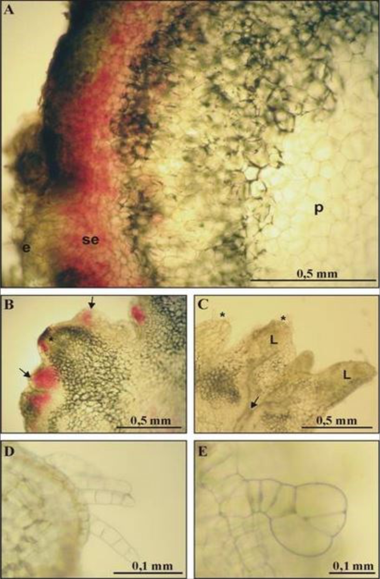 |
Fig. 1: Histological structure of yacon (Smalanthus sonchifolius) tissue obtained on leaves on MS medium supplemented with 1mg l-1 TDZ and 1 mg l-1 NAA; Fig. 1a and b: red external layers of cells containing chlorophyll and centrally located, colourless parenchyma cells Fig. 1b and c: organogenesis process on the different level of advancement on the surface of callus tissue Fig. 1d: tissues of multicellular trichomes within the epidermis of a cover character Fig. 1e: tissues of multicellular trichomes within the epidermis of a glandular character e: epidermal cells, se: subepidermal cells, p: parenchymal cells, L: leaf development |
DISCUSSION
Estrella and Lazarte14, used with a success 80% ethanol and 1% calcium hypochlorite with 0.05% Tween 20 for 20 min. to sterilize shoots collected from plants grown in the field. In this experiment, a similar combination two-step disinfection method was too drastic for initial explants, resulting in low survival and poor regeneration capacity.
Mogor et al.16 used buds sprouting from tuberous roots as initial explants of yacon. They disinfected them with 20% sodium hypochlorite for 20 and 40 min. However, a high frequency of bacterial and fungal infections was observed for those explants. To initiate in vitro cultures of yacon, the various explant types were used successfully: apical buds15, lateral buds14,16 shoot and root fragments17, nodes, leaves and their fragments18. Matsubara et al.15 obtained single plants from growth apical buds, two shoots from each node and callus, shoots or roots from leaf fragments. Estrella and Lazart14 reported shoots development from axillary buds. All explants tested in this research demonstrated proliferation ability.
The manner and direction of shoot development depended not only on the explant type, its structure or origin but also on the applied medium and PGRs composition. MS medium was the most often used medium for yacon micropropagation. Paterson et al.19 used MS medium for the shoot induction from callus and leaf explants, which was also applied in our experiments. Matsubara et al.15 used MS medium supplemented with 0.01 mg L-1 NAA and 0.01 mg L-1 BA for apex buds. The MS medium with the addition of the same growth substances was applied for nodes. MS medium proved to be the most effective. Authors applied MS medium supplemented with 2,4-D and BA in various concentrations for leaf fragments, shoots and roots. The most effective proved to be MS medium supplemented with 0.1 mg L-1 2,4-D and BA. The same medium was used by Niwa et al.18 for leaf fragments. Callus formation was observed most frequently on this medium. Callus production on MS medium containing 0.1 mg L-1 2,4-D and 1 mg L-1 BA was reported by Matsubara et al.15. These authors observed sporadic adventitious embryos formation after transferring callus on MS medium supplemented with 0.1 mg L-1 BA. Estrella and Lazarte14 used MS medium supplemented with IBA and BA and axillary buds to initiate in vitro cultures of yacon. In their studies, the most effective PGRs combination proved to be the 0.1 mg L-1 of IBA and 2 mg L-1 of BA. Niwa et al.18 achieved shoot organogenesis from callus (obtained on leaf fragments) on HaR medium supplemented with 1 mg L-1 BA and 0.1 mg L-1 GA3. Plants micro propagated on this medium were submitted to AFLP analysis which revealed the genetic variation.
Knowing the mutagenic impact of 2,4-D on micropropagation yacon plants, described by Niwa et al.18, this growth regulator was excluded from this experiments. Various types of cytokinin were used for inducing regeneration from leaves. The TDZ was included in this experiment because there is a lot of information about its positive effects on induction, shoot regeneration and multiplication from different explants of various species, including leaves20.
Singh and Dwivedi21 applied various cytokinins during conducting studies on the multiplication of Stevia reubadiana. Authors discovered that all cytokinins influenced shoot formation, but the most effective was TDZ. Brijwal et al.22 received the biggest number of shoots from callus collected from leaves of Berberis aristata using WP medium (Woody Plant) with the addition of 0.02 mg L-1 TDZ. Siddique et al.23 used BA, Kin and TDZ to induce shoot formation from buds during the research on micropropagation of Cassia angustifolia. The best results were obtained during the culture on the medium supplemented with 1 mg L-1 TDZ. Slesak et al.24 reported the efficient shoot regeneration from root fragments of Rumex thyrsiflorus on MS medium supplemented with 0.5 mg L-1 TDZ. High regeneration frequencies of adventitious shoots from bulb scale and flower stem in the presence of TDZ was reported for bulbous plants such as Tulipa gesneriana and Fritllaria meleagris, respectively25,26. Tsai et al.27 elaborated the method of intensive multiplication of Salvia multioryzae from leaf explants. The largest number of shoots was obtained by direct organogenesis with the use of a medium with 0.1–0.5 mg L-1 TDZ. Nieves et al.28 obtained the greatest number of Calanchoe blossfeldiana shoots from node explants on the medium supplemented with 0.2 mg L-1 TDZ.
In this study, the growth of a specific tissue was observed when leaves and their fragments were cultured in the presence of TDZ and NAA, both at the concentration of 1 mg L-1. That was similar to the tissue described by Hong et al.29 in orchid Paphiopedillum. The adventitious shoot regeneration was induced after transferring this tissue on a medium containing 5 mg L-1 BA and 1 mg L-1 IBA. Anatomical analysis of those tissues confirmed the presence of adcentitious shoot buds. In the current opinion, it is a great achievement that we managed to stimulate leaves and their fragments to generate the numerous buds using TDZin the combination with NAA, without the use of 2,4-D. Matsubara15 reported that yacon shoots undergo the rooting process with ease. Estrella and Lazaarte14 obtained complete plants from axillary buds cultured on a medium containing 0.02 mg L-1 BA and 0.1 mg L-1 IBA. Niwa et al.18 obtained rooted shoots on ½ MS media supplemented with 0.1 mg L-1 NAA. In other studies, yacon shoots were rooted successfully on MS medium containing 0.1 mg L-1 BA13.
Estrella and Lazarte14 reported that only 40% of plants adapted to ex vitro conditions. According to Hamada et al.13, yacon plants adapted easily in the aired substrate under plastic tunnel within two weeks. Matsubara15 also presented that yacon plants undergo easily the acclimatization process. Also, in this experiment, it was discovered that yacon plants adapted quickly and easily to external conditions. Conducted studies allowed to gain the skill of controlling the process of organogenesis in yacon by using various explants and selecting proper combinations of growth regulators such as auxin and cytokinin and their concentrations. The result of this study is the elaboration of technology of intensive multiplication of yacon.
CONCLUSION
Results of conducted studies allowed elaborating an efficient technology of multiplication of yacon in vitro conditions, in which each stage of micropropagation was optimized. The optimal explants for the culture induction are shoot tips and lateral buds from which after the surface sterilization with the I method, 1 mm long growth tips were isolated. The best for the culture induction proved to be the MS medium supplemented with 2 mg L-1 kinetin, 1 mg L-1 IAA and also the medium with the addition of 5 mg L-1 BA, 1 mg L-1 IBA. On both these media, the process of callusgenesis, organogenesis and also shoot multiplication occur. On the first medium, the multiplication of small shoots is lower, but formed shoots are ready for the rooting process, whereas, on the second medium, the multiplication of small shoots is higher. In the conducted experiment, besides the shoots, a proliferating tissue was obtained on these media, which multiplied the best on MS medium supplemented with 1 mg L-1 TDZ and 1 mg L-1 NAA. Shoots designated for rooting should be placed on MS ½ or ¼ media, containing 20 mg L-1 sucrose and 1 mg L-1 NAA, which showed the 100% effectiveness of rhizogenesis in the conducted experiment.
SIGNIFICANCE STATEMENT
Conducted studies showed that Yacon is a plant with a great potential for multiplication in the tissue culture. Obtained results are the basis for elaborating a technology for the production of plant tissue in bioreactors. Thus, there will be a possibility of the wider study and usage of the bioactive substances discovered in this plant, in particular the anticarcinogenic components.
REFERENCES
- Valentova, K., A. Moncion, I. de Waziers and J. Ulrichova, 2004. The effect of Smallanthus sonchifolius leaf extracts on rat hepatic metabolism. Cell Biol. Toxicol., 20: 109-120.
- Santana, I. and M.H. Cardoso, 2008. Raiz tuberosa de yacon (Smallanthus sonchifolius): potencialidade de cultivo, aspectos tecnológicos e nutricionais. Ciência Rural, 38: 898-905.
- Lago, A., S.M. Godden, R. Bey, P.L. Ruegg and K. Leslie, 2011. The selective treatment of clinical mastitis based on on-farm culture results: i. effects on antibiotic use, milk withholding time, and short-term clinical and bacteriological outcomes. J. Dairy Sci., 94: 4441-4456.
- Cazetta, M.L., P.M.M. Martins, R. Monti and J. Contiero, 2005. Yacon (Polymnia sanchifolia) extract as a substrate to produce inulinase by Kluyveromyces marxianus var. bulgaricus. J. Food Eng., 66: 301-305.
- Milella, L., G. Martelli, J. Salava, E. Fernandez, J. Ovesna and I. Greco, 2011. Total phenolic content, RAPDs, AFLPs and morphological traits for the analysis of variability in Smalanthus sonchifolius. Genet. Resources Crop Evol., 58: 545-551.
- Lachman, J., E.C. Fernández and M. Orsák, 2011. Yacon [Smallanthus sonchifolia (poepp. et endl.) h. robinson] chemical composition and use – a review. Plant, Soil Environ., 49: 283-290.
- Passos, L.M.L. and Y.K. Park, 2003. Frutooligossacarídeos: implicações na saúde humana e utilização em alimentos. Ciênc. Rural, 33: 385-390.
- Aybar, M.J., A.N. Sanchez Riera, A. Grau and S.S. Sanchez, 2001. Hypoglycemic effect of the water extract of Smallantus sonchifolius (yacon) leaves in normal and diabetic rats. J. Ethnopharmacol., 74: 125-132.
- Simonovska, B., I. Vovk, S. Andrenšek, K. Valentová and J. Ulrichová, 2003. Investigation of phenolic acids in yacon (Smallanthus sonchifolius) leaves and tubers. J. Chromatogr. A, 1016: 89-98.
- Corrêa, C.M., G.N. de Oliveira, L.V. Astarita and E.R. Santarém, 2009. Plant regeneration through somatic embryogenesis of yacón [Smallanthus sonchifolius (poepp. and endl.) h. robinson]. Braz. Arch. Biol. Technol., 52: 549-554.
- Li, J., J. Liu, H. Lan, M. Zheng and T. Rong, 2009. Gc-ms analysis of the chemical constituents of the essential oil from the leaves of yacon (Smallanthus sonchifolia). Front. Agric. China, 3: 40-42.
- Mansilla, R., C. López, M. Flores and R. Espejo, 2010. Reproductive biology syudy in five accessions of Smallanthus sonchifolius (Poepp. and Endl.), Robinson Ecol. Appl., 9: 167-175.
- Hamada, M., T. Hosoki and Y. Kusabiraki, 1990. Mass-propagation of yacon (Polymnia sonchifolia) by repeated node culture.. Plant. Tissue. Cult. Lett., 7: 35-37.
- Estrella, J.E. and J.E. Lazarte, 1994. In vitro propagation of ji?cama (Polymnia sonchifolia poeppig & endlicher): a neglected andean crop. Hortic. Sci., 29: 331-337.
- Matsubara, S., 1997. Micropropagation of Polymnia sonchifolia (yacon). Biotechnol. Agric. For., 39: 150-159.
- Mogor, G., A.F. Mogor and G.P.P. Lima, 2003. Bud source, asepsis and benzylaminopurine (bap) effect on yacon (Polymnia sonchifolia) micropropagation. Acta Hortic., 597: 311-314.
- Murashige, T. and F. Skoog, 1962. A revised medium for rapid growth and bio assays with tobacco tissue cultures. Physiol. Planta., 15: 473-497.
- Niwa, M., T. Arai, K. Fujita, W. Marubashi, E. Inoue and T. Tsukihashi, 2002. Plant regeneration through leaf culture of yacon. J. Jpn. Soc. Hortic. Sci., 71: 561-567.
- Paterson, K.E. and N.P. Everett, 1985. Regeneration of helianthus annuus inbred plants from callus. Plant Sci., 42: 125-132.
- Ernst, R., 1994. Effects of thidiazuron on in vitro propagation of Phalaenopsis and Doritaenopsis (Orchidaceae). Plant. Cell. Tiss. Org., 39: 273-275.
- Singh, P. and P. Dwivedi, 2013. Two-stage culture procedure using thidiazuron for efficient micropropagation of Stevia rebaudiana, an anti-diabetic medicinal herb. 3 Biotech, 4: 431-437.
- Brijwal, L., A. Pandey and S. Tamta, 2015. In vitro propagation of the endangered species Berberis aristata DC. via leaf-derived callus. in vitro Cell. Dev. Biol. Plant, 51: 637-647.
- Siddique, I., N.A.W. Bukhari, K. Perveen and I. Siddiqui, 2015. Influence of plant growth regulators on in vitro shoot multiplication and plantlet formation in Cassia angustifolia vahl. Braz. Arch. Biol. Technol., 58: 686-691.
- Slesak, H., G. Góralski, D. Kwolek, K. Dziedzic and A. Grabowska-Joachimiak, 2015. Male adventitious roots of rumex thyrsiflorus fingerh. as a source of genetically stable micropropagated plantlets. Plant Cell, Tiss. Org. Cult., 123: 193-203.
- Podwyszy?ska, M. and A. Marasek, 2011. Effects of thidiazuron and paclobutrazol on regeneration potential of tulip flower stalk explants in vitro and subsequent shoot multiplication. Acta Soc. Bot. Pol., 72: 181-190.
- Petric, M., A. Subotic, S. Jevremovic, M. Trifunovic-Momcilov, V. Tadic, M. Grujic, Z. Vujcic, 2015. Esterase and peroxidase isoforms in different stages of morphogenesis in Fritillaria meleagris L. in bulb-scale culture. C.R. Biol., 338: 793-802.
- Tsai, K.L., E.G. Chen and J.T. Chen, 2015. Thidiazuron-induced efficient propagation of Salvia miltiorrhiza through in vitro organogenesis and medicinal constituents of regenerated plants. Acta Physiol. Plant., 38: 29-39.
- Nieves, M.C., E.T. Aspuria, E.C. Bernardo and M.A.D. Tayangona, 2016. Growth response of in vitro-derived nodal sections of Kalanchoe blossfeldiana poellnitz as influenced by benzylaminopurine, thidiazuron and paclobutrazol. Asia Life. Sci.,, 25: 207-220.
- Hong, P.I., J.T. Chen and W.C. Chang, 2008. Plant regeneration via protocorm-like body formation and shoot multiplication from seed-derived callus of a maudiae type slipper orchid. Acta Physiologiae Plantarum, 30: 755-759.
How to Cite this paper?
APA-7 Style
Kiszczak,
W., Kowalska,
U., Burian,
M., Glinska,
S., Górecka,
K. (2021). Influence of Growth Regulators on Micropropagation System of Medicinal Plant Yacon (Smallanthus sonchifolius (Poepp.) H. Rob.). Asian Journal of Emerging Research, 3(1), 44-48. https://doi.org/10.3923/ajerpk.2021.44.48
ACS Style
Kiszczak,
W.; Kowalska,
U.; Burian,
M.; Glinska,
S.; Górecka,
K. Influence of Growth Regulators on Micropropagation System of Medicinal Plant Yacon (Smallanthus sonchifolius (Poepp.) H. Rob.). Asian J. Emerg. Res 2021, 3, 44-48. https://doi.org/10.3923/ajerpk.2021.44.48
AMA Style
Kiszczak
W, Kowalska
U, Burian
M, Glinska
S, Górecka
K. Influence of Growth Regulators on Micropropagation System of Medicinal Plant Yacon (Smallanthus sonchifolius (Poepp.) H. Rob.). Asian Journal of Emerging Research. 2021; 3(1): 44-48. https://doi.org/10.3923/ajerpk.2021.44.48
Chicago/Turabian Style
Kiszczak, Waldemar, Urszula Kowalska, Maria Burian, Sława Glinska, and Krystyna Górecka.
2021. "Influence of Growth Regulators on Micropropagation System of Medicinal Plant Yacon (Smallanthus sonchifolius (Poepp.) H. Rob.)" Asian Journal of Emerging Research 3, no. 1: 44-48. https://doi.org/10.3923/ajerpk.2021.44.48

This work is licensed under a Creative Commons Attribution 4.0 International License.
Division of Scientific Publishing
ACE College for Women
Faisalabad-38090, Pakistan
ISSN: 2663-4988 / 2664-5211
This work is licensed under a Creative Commons Attribution 4.0 International License.