
A Few Inflammatory Markers Linked to Diabetic Nephropathy
-
Kaiser Jamil


Department of Genetics, Bhagwan Mahavir Medical Research Centre, 10-1-1, Mahavir Marg, 500004, Hyderabad, TS, India
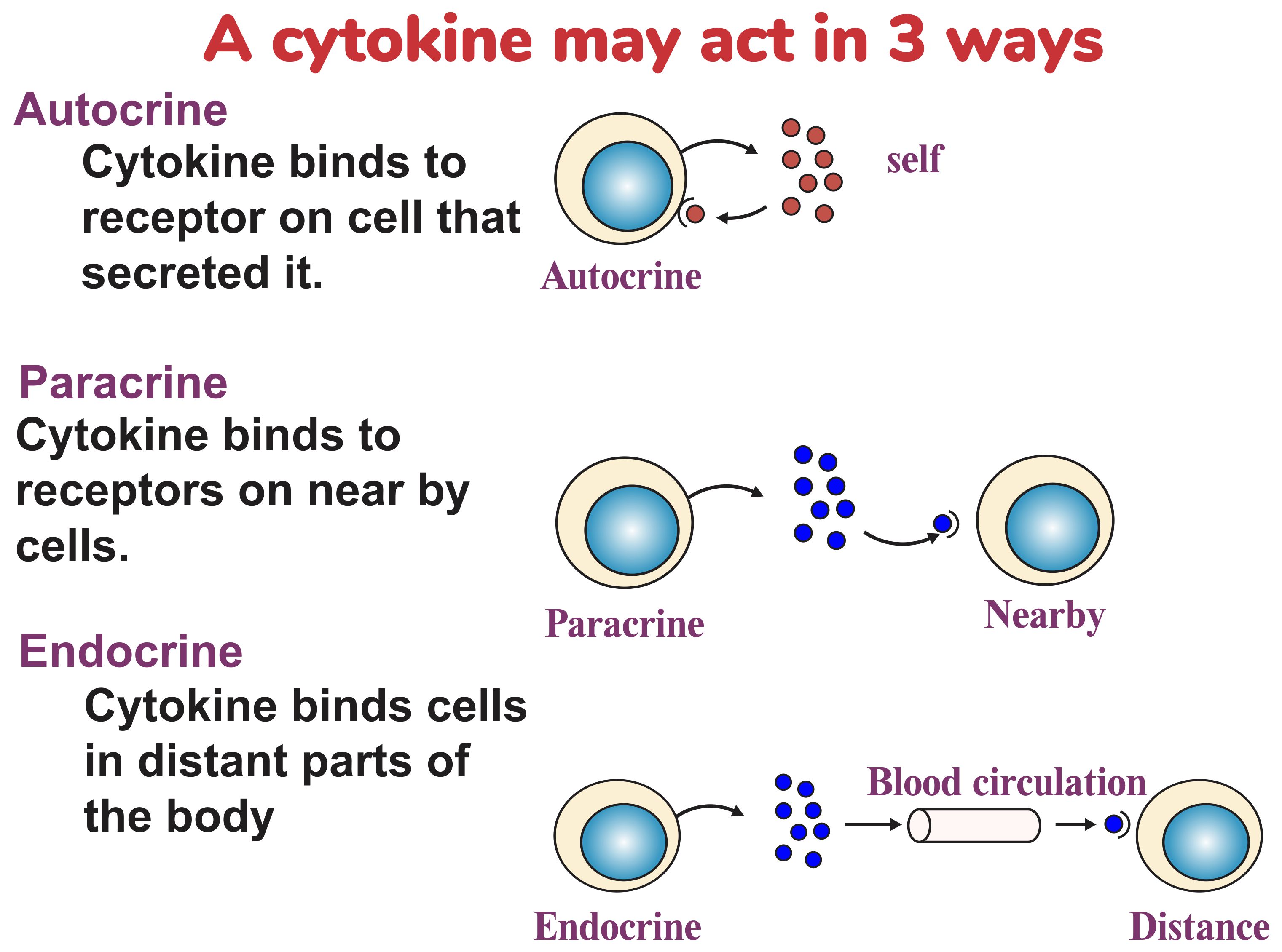
Several articles have been published in the last decade on inflammatory markers in several diseases; here I am looking at the most popular inflammatory markers of Diabetes mellitus. These are Cytokines which include chemokines, interferons, interleukins, lymphokines, and tumour necrosis factors. These are signalling molecules that mediate and regulate immunity, inflammation and haematopoiesis. Generally these are low molecular weight proteins, peptides or glycoproteins secreted by a broad range of cells, that include immune cells like macrophages, B lymphocytes, T lymphocytes and mast cells as well as endothelial cells, fibroblasts and various stromal cells. They are involved in endocrine, paracrine and autocrine signalling. Controlling cell populations is on one of their jobs, besides being important in immune system where they modulate the balance between humoral and cell-based immune responses acting through the receptors. They are important in the host responses to infection, inflammation, trauma, cancer, reproduction etc, and play an important role in health and disease. A given cytokine may be produced by more than one type of cell.
Few publications of 1991 suggested for the first time that inflammatory cytokines may participate in the pathogenesis of diabetic nephropathy. Today, it is known that among inflammatory cytokines, IL-1, IL-6, IL-18 and TNF- were found to be closely relevant to the development of diabetic nephropathy, with diverse actions potentially involved in the development of complications. Literature also reveals that cytokines are involved in micro-angiopathic complications of Diabetic Mellitus. Based on this knowledge some cytokines such as Interleukins were classified as anti-inflammatory and pro-inflammatory molecules due to their physiological actions. It is known that Interleukin-1 promotes an increase of adhesion molecules in glomerular endothelium, further the expression of these molecules is also seen in other kidney structures. Interestingly, IL-1 stimulates hyaluronan synthesis, in diabetic patients which leads to cell proliferation, and subsequently leading to the development of diabetic nephropathy in DM.
Interleukins (IL-1, IL-6 and IL18):
Time and again experimental models have demonstrated increased renal expression of interleukins in diabetic nephropathy which is related to subsequent expression of chemotactic factors and adhesion molecules. IL-1 is known to deregulate the production of hyaluronan by renal proximal tubular epithelial cells. Hence it was established by authors that patients with type 2 DN had higher levels of IL-6 in their serum than with diabetic patients without nephropathy. Such finding surely suggests the role played by cytokine in the pathogenesis of DN.It is a potent cytokine that induces IFN-γ, which in turn induces functional chemokine receptor expression in human mesangial cells. Patients with DN have shown increased levels of IL-18 in serum and urine with an independent relationship between these parameters and urinary albumin excretion. Moreover, some findings from a longitudinal study showed that serum and urinary concentrations of this cytokine were directly correlated with albumin excretion rate, as well as with changes in albuminuria during the follow-up period.
TNF-α:
TNF alpha is a pleotropic inflammatory cytokine mainly produced by monocytes, macrophages and T-cells. It is known to act on the renal cells in a number of ways such as by activating the second messenger system, enzymes involved in the synthesis of other inflammatory mediators, transcription factors, cell adhesion molecules, synthesis of cytokines, growth factors, receptors, MHC proteins and acute phase proteins. The various biologic activities of TNF-α resulted in diverse effects along with a significant role in the development of renal damage in diabetes. Also due to the cytotoxic effects of TNF-α, it can induce direct renal injury and higher levels of TNF-α could be found in the urine and serum of DN patients than in diabetes patients with no nephropathy. The way in which TNF-α can induce such a variety of effects on the renal structure could be in a paracrine or autocrine manner. Further, TNF-α can have a direct effect on protein permeability barrier of the Glomeruli, independent of the effects of recruited inflammatory cells which were synthesized in the renal membrane.
Several recent studies suggest that the inflammatory processes are implicated in the onset of diabetes and the progression of the complication. Through the activation of NF-κB pathway, the development of insulin resistance in type 2 diabetes was found to be associated with the release of inflammatory cytokines and other mediators from adipose cells. Obesity, hyperglycaemia, a high fat diet, PKC activation and oxidative stress are the other factors that activate the NF-κB pathway with the development of insulin resistance. A well-described pathway underlying the development of diabetic vascular complications is via activation of Protein kinase- (PKC) isoforms, PKC-b in-particular. At the level of individual tissues, activation of PKC-b is associated with the development and/or progression of microvascular and macrovascular complications. Hyperglycaemia is the major risk factor for the development of micro-vascular diabetic irregularities such as retinopathy, neuropathy and nephropathy. Although evidence suggests that the disease is associated with the increase in inflammatory markers, the major risk factor for its development in diabetes is still unclear.
However one fact remains clear that Tumor necrosis factor-alpha (TNF-α) is one of the proinflammatory cytokines responsible for inflammation, immunity, and cellular homeostasis.
TNF-α may mediate excitotoxic neurodegeneration by inhibiting the activity of the insulin-like growth factor I (IGF-I), a neuroprotective peptide that stresses the interaction between neurotrophic and inflammatory factors. TNF-α also plays a critical role in inflammation and is a key regulator of the microenvironment. Hence could also be linked to the pathophysiology of T2DN.
How to Cite this paper?
APA-7 Style
Jamil,
K. (2019). A Few Inflammatory Markers Linked to Diabetic Nephropathy . Asian Journal of Emerging Research, 1(2), 51-53. https://doi.org/10.3923/AJERPK.2019.51.53
ACS Style
Jamil,
K. A Few Inflammatory Markers Linked to Diabetic Nephropathy . Asian J. Emerg. Res 2019, 1, 51-53. https://doi.org/10.3923/AJERPK.2019.51.53
AMA Style
Jamil
K. A Few Inflammatory Markers Linked to Diabetic Nephropathy . Asian Journal of Emerging Research. 2019; 1(2): 51-53. https://doi.org/10.3923/AJERPK.2019.51.53
Chicago/Turabian Style
Jamil, Kaiser.
2019. "A Few Inflammatory Markers Linked to Diabetic Nephropathy " Asian Journal of Emerging Research 1, no. 2: 51-53. https://doi.org/10.3923/AJERPK.2019.51.53

This work is licensed under a Creative Commons Attribution 4.0 International License.