
Anti-Ulcerogenic Efficacy of Leaf Fractions of Argemone mexicana Against Indomethacin Induced-Ulceration in Rats
-
O.A. Idowu


Department of Biochemistry, College of Natural and Applied Sciences, Oduduwa University, P.M.B 5533 Ile-Ife, Osun State, Nigeria
O.A. SaliuDepartment of Environmental Health, Faculty of Health Sciences, National Open University, Abuja, Nigeria
N.C. FakoredeDepartment of Microbiology, College of Natural and Applied Sciences, Oduduwa University, P.M.B 5533 Ile-Ife, Osun State, Nigeria
R.O. AriseDepartment of Biochemistry, University of Ilorin, Ilorin Kwara State, P.M.B 1515, Ilorin, Nigeria
| Received 17 Feb, 2021 |
Accepted 12 Apr, 2021 |
Published 15 Jun, 2021 |
Background and Objective: Peptic ulcer disease still remains a burden in under-developed and developing countries. Argemone mexicana is a plant locally used in folk medicine of Nigeria to treat peptic ulcer disease. Therefore this study aimed to investigate the anti-ulcer activity of solvent-partitioned fractions of Argemone mexicana leaves in indomethacin ulcerated rats. Materials and Methods: Dried leaves of A. mexicana were extracted with ethanol and the crude extract was subjected to solvent-partitioning using n-hexane, ethylacetate and butanol. Phytochemical screening of the partitioned fractions was carried out using standard methods. A total of 36 rats were randomized into six groups of six rats each. Group 1 serve as the control and received distilled water only, Group 2 serve as ulcerated group, Groups 3 serve as ulcerated group treated with 20 mg kg-1 b.wt., Omeprazole while Groups 4-6 were ulcerated rats treated with 200 mg kg-1 b.wt., of ethylacetate, butanol and n-hexane fractions respectively. The rats were ulcerated by single dose of indomethacin (25 mg kg-1 b.wt.,) administered orally followed by treatment with the fractions. Ulcer indices, antioxidant enzymes status, levels of glutathione and lipid peroxidation were assessed. Results: The results revealed the presence of alkaloids and flavonoids only in ethylacteate and butanol fractions. A significant increase (p<0.05) in gastric acid, volume, malondialdehyde concentration, pepsin and H+/K+ ATPase activities was observed in the ulcerated rats. Significant decrease (p<0.05) was observed in gastric mucus content, glycoprotein concentration and antioxidant activities in ulcerated rats. The ulcer indices were attenuated and the antioxidant status was improved in rats treated with 200 mg kg-1 b.wt., of ethylacetate and butanol fractions. Conclusion: The ethylacetate fraction demonstrated a better efficacy which may be attributed to presence of flavonoids thus suggesting Argemone mexicana as an alternative therapy for treating ulcer.
INTRODUCTION
Peptic Ulcer Disease (PUD) is still a burden affecting most of the populace in under-developed and developing countries. The gastrointestinal tract (GIT) plays a significant role in digestion and absorption of food materials in the living system however, disturbances in physiological functions of the GIT among which are alterations in acid secretion and osmotic effect can result to several pathological conditions of the gastrointestinal tract such as diarrhea, appendicitis, cancer and ulcer1. An ulcer is an open sore that develops in the mucosa lining the GIT. A conglomerate of ulcer that develops in the lining mucosa of the oesophagus (oesophagael ulcer), stomach (gastric ulcer), duodenum (duodenal ulcer) and meckel’s diverticulum (meckel’s diverticulum ulcer) of the gastrointestinal tract are referred as gastrointestinal tract ulcers. Among these ulcers, gastric and/or duodenal ulcers most often referred as peptic ulcer disease are the most common among the populace. Peptic ulcer usually results from an imbalance between aggressive/offensive factors (acid, pepsin) and defensive factors (prostaglandin, mucus, glycoprotein, bicarbonate and antioxidant enzymes) of the stomach especially when the aggressive/offensive factors overwhelm the defensive factors2. Such imbalance may be induced by Helicobacter pylori infection, excessive consumption of Non-Steroidal Anti-inflammatory Drugs (NSAIDs), smoking, alcohol consumption and diet2 as well as physiological (oxidative) and psychological stress3. The NSAIDs e.g. indomethacin induce peptic ulcer by inhibiting cyclooxygenase, an enzyme responsible for the biosynthesis of prostaglandin which is a major biomolecule that enhance the defense barrier of gastric mucosal against the damaging effect of gastric acid by stimulating mucus secretion, glycoprotein and other defensive factors. Indomethacin and other NSAIDs bind to the active site of cyclooxygenase and acetylate its serine residue thereby causing the inactivation of the enzyme. The inhibition of this enzyme finally suppresses the production of prostaglandin and subsequently impedes the secretion of mucus and result in ulceration. Normally, an equilibrium exist between the acid (HCl) secreted by the stomach which help in the digestion of food and the gastric mucosal defensive factors however, when an imbalance between these two is induced by any of the exogenous agents (e.g. NSAIDs, H. pylori, stress etc.), the mucosal becomes irritated by the bathing of the acid which weakens it and then result to ulcer. Symptoms of peptic ulcer include abdominal pain, nausea, vomiting, loss of appetite and weight loss. Ulcers could get complicated and show symptoms of bleeding and perforation which ulcer may require surgery. A number of drugs currently available for treating gastrointestinal tract ulcers are histamine-receptor blockers (e.g. ranitidine, cimetidine), proton-pump blockers (e.g. omeprazole, pantoprazole), prostaglandins analogues (e.g. misoprostol, sucralfate) antibiotics (e.g. clarithromycin amoxicillin, metronidazole), antacids (Aluminum hydroxide, Sodium hydroxide), synthetic antioxidants (e.g. dubinol, sodium benzoate) etc. Although these conventional drugs have been well characterized but the adverse effects they produce such as headache, male hormone disturbances4, pneumonia, osteoporosis, vitamin B12 malabsorption5, hematopoietic changes and drug-drug interaction highlight the need for better treatment modalities of gastrointestinal tract ulceration of any kind1. Recent studies found that different substances from plant sources not only afford gastro protection and accelerate ulcer healing but are also safe to consume6. There are various plant used as decoction, concoction, or even as food additive or supplement to combat gastrointestinal ulceration in folk medicine of many countries6. In Nigeria, a West Africa country, ethno botanical survey revealed the use of Argemone mexicana as treatment for peptic ulcer. A. mexicana belongs to the family Papaveraceae. It is an annual herb commonly found in tropical and subtropical regions of the world among which are Indian, Mexico and Nigeria7. It leaves are estipulate, sessile, alternate, deeply lobed, cauline with unicostate reticulate venation with thorny margins8. Traditionally, in Mali, Burkina Faso and Nigeria, the decoction leaves of the plant are used to treat cough, uncomplicated malaria, liver disease and ulcer9. However, there is need to provide substantial scientific information to validate its purported traditional claims for treating ulcer. This study therefore aimed to investigate the effects of different solvent partitioned fractions of Argemone mexicana leaves on acid secretory parameters and antioxidant status of indomethacin-induced ulcerated rats.
MATERIALS AND METHODS
Study area: This study was carried out between June and December, 2015 at the Department of Biochemistry, University of Ilorin, Nigeria.
Collection of plant materials and authentication: Fresh leaves of A. mexicana were collected from Saki Township, Oyo State, Nigeria. They were identified and authenticated at the Department of Botany, University of Ilorin, Kwara State, Nigeria. A voucher specimen number of the plant (UIH0011171) was deposited in the Herbarium of the Department.
Preparation of extract: The fresh leaves were rinsed with distilled water and air-dried for 14 days in the laboratory at room temperature (25oC) to prevent loss of bioactive agents due to irradiation. The dried leaves were ground to powder form using mechanical blender (Mazeda Mill, MT 4100, Japan). Almost 500 g of the powder was macerated with absolute ethanol (2 L) for 72 h using cold extraction method. The crude extract was filtered using Whatman No 1 filter paper. The extraction process was repeated twice with the marc and the three filtrates obtained were combined and concentrated using a rotary evaporator. The crude extract was dissolved in distilled (10% w/v) water and was subsequently subjected to successive solvent partitioning in order of polarity using n-hexane, ethyl acetate and butanol. Each fraction obtained was filtered, concentrated and the resulting residues were reconstituted in distilled water to give the required dose of 200 mg kg-1 b.wt.
Phytochemical profiling: The phytochemical screening for each of the fractions was carried out using standard procedures10,11,12.
Experimental animals: A total of thirty-six (36) albino rats (Rattus norvegicus) of both sexes weighing 162±1.45 g were obtained from the Animal Holding Unit, Biochemistry Department University of Ilorin, Kwara State, Nigeria. They were housed under standard conditions temperature: 22±3oC; photoperiod: 12 h light and 12 h dark; humidity: 40–45% with free access to rat pellets (Premier Feed Mills Company Limited, Ibadan, Nigeria) and tap water ad libitum. Full committee approval was given by the University of Ilorin, Nigeria ethical review committee (UERC) with protocol approval number of UERC/ASN/2015/120.
Chemicals: Omeprazole and indomethacin were purchased from Pauco Pharmaceutical (Anambra, Nigeria). Trichloroacetic acid (TCA), 5,5′-dithiobis-(-2-nitrobenzoic acid) (DTNB) and thiobarbituric acid (TBA), potassium ferricyanide, nicotinamide adenine dinucleotide (NADH), ferric chloride were products of Sigma-Aldrich Inc. (St. Louis, MO, USA). Glutathione peroxidase (GSH-px), glutathione reductase (GSH-Red), glutathione transferase (GST) and superoxide dismutase were purchased from Randox Laboratories (Antrim, United Kingdom). All other chemicals used in this research are of analytical grade and purchased commercially.
Experimental procedure: The animals were assigned into six groups (I-VI) consisting of six rats each. Group I received distilled water only and served as the control. Group II are rats ulcerated with indomethacin but receive no chemical intervention. Group III are ulcerated rats with medical intervention of 20 mg kg-1 b.wt., omeprazole as reference drug while groups IV-VI are ulcerated rats that received medical intervention of 200 mg kg-1 b.wt., of ethylacetate, n-hexane and butanol partitioned fractions of A. mexicana respectively. The rats were fasted for 24 h before the commencement of the experiment but had access to water until 6 h before the experiment after which they were ulcerated by single oral administration of 25 mg kg-1 b.wt., indomethacin. The rats had access to water till the next two days for the establishment of ulceration after which administration of the vehicle, reference drug and the plant fractions began on the third day and lasted for 7 days. The animals were again fasted for 12 h to ensure complete gastric emptying and a steady state gastric acid secretion after which they were anesthetized under diethyl ether. The stomach of the rats was ligated at both openings of the lower esophageal sphincter and pyloric sphincter and injected with 3 mL of distilled water to collect the gastric juice6 which was used for biochemical analysis.
Preparation of tissue homogenates: The stomach and duodenum were excised and placed in cold 0.25 M sucrose solution to maintain the integrity of the organs. The organs were separately homogenized in ice-cold 0.25 M sucrose solution. The homogenates were appropriately diluted (1:5w/v) with sucrose and centrifuged at 1000xg for 10 min. The supernatant were aspirated with Pasteur pipette into sample bottles and refrigerated until further analysis.
Ulcer index determination: After the collection of the gastric juice, the stomach was ligated along the greater curvature and rinsed slowly with normal saline. It was then stretched out on a plane paper and examined macroscopically with a hand lens (x20) for gastric erosion. For the ulcer index, the length (mm) and width (mm) of ulcer on the gastric mucosa was measured using a ruler using the procedure of Gregory et al.13. The Ulcer Index (UI) was then calculated in mm2 using the expression:
Ulcer Index (UI) = Length (mm) x Breadth (mm) of lesion
Determination of ulcer indices in the gastric juice: The gastric juice collected was centrifuged at 850 x g for 10 min. The volume of the supernatant was measured using a graduated measuring cylinder and was taken as the volume of the gastric juice6. The pH was measured by placing the electrode of a digital pH meter into the gastric juice supernatant. The procedure of Maity et al.14 was used to determine the gastric acidity.
Pepsin activity in gastric juice: The method described by Hirohashi et al.15 was used to determine pepsin activity in the gastric juice. Briefly, 1.0 mL of gastric juice supernatant and 5 mL of buffer solution (0.2N sodium citrate and 0.2N HCl pH 1.2 in ratio 1:4) in different test tubes were incubated at 37oC for 30 min. Then pepsin was allowed to react with 2 mL bovine serum albumin (10 mg) and test tubes re-incubated at 37oC for 30 min. The unreacted protein of bovine serum albumin was detected by the addition of 1.0 mL of Biuret reagent and absorbance was read at 546 nm after 30 min against a reagent blank. The pepsin activity was determined from standard protein curve.
Determination of glycoprotein concentration: Glycoprotein concentration was determined from the total carbohydrate and protein contents expressed as ratio of Total Carbohydrates (TC) to Total Proteins (TP).
Total carbohydrates: The total carbohydrate in the gastric juice was determined by the method of Nair16. Briefly, to 0.15 mL gastric juice and to blank containing 0.15 mL of distilled water in a test tube, 1 mL of 5% phenol was added separately and mixed thoroughly. The 5 mL of 96% H2SO4 was added and again mixed slowly. After 10 min, the test tubes were shaken and placed in water bath kept at 20°C for 20 min. The optical density of the developed yellow orange chromophore was read in a UV spectrophotometer at 482 nm. Several concentrations of glucose standard solution were run to prepare a standard curve. Total carbohydrates were expressed in terms of micrograms per milliliter liberated in gastric juice.
Total protein: The procedure described by Lowry et al.17 was used for the determination of total protein content in the gastric juice.
The glycoprotein concentration was thereby expressed as the ratio of total carbohydrates and protein content below.
$$ \text { Glycoprotein }=\frac{\text { Total Carbohydrate }}{\text { Total Protein }} $$Determination of biochemical parameters in stomach and duodenum
H+/ K+ -ATPase activity: The H+/K+-ATPase activity in the gastric mucosa was assayed for by the method of Reyes-Chilpa et al.18.
Adherent gastric mucus content: The adherent gastric mucus content in the stomach was determined by the procedure of AlRashdi et al.19. Glandular segments of the stomach was removed and weighed. Each segment was immediately placed into 10 mL 0.1% w/v alcian blue solution (in 10 mL of 0.16 M sucrose solution, buffered with 0.05 M sodium acetate pH 5). After immersion for 2 h, excess dye was removed by successively rinsing twice with 10 mL of 0.25 M sucrose, first for 15 min then later for 45 min. The dye complexed with stomach wall mucus was extracted with 10 mL of 0.5 M MgCl2 with intermittent shaking for 1 min at 30 min intervals for 2 h. The 4 mL of the alcian blue extract was added with an equal volume of diethyl ether and shaken vigorously. The emulsion obtained was span at 725 x g for 10 min and absorbance of an aqueous layer formed was read at 580 nm. Values were compared with alcian blue concentration standard curve.
Reduced glutathione concentration: The reduced glutathione concentration was determined in the stomach and duodenum using the procedure of Ellman20.
Antioxidant enzymes: Superoxide dismutase (SOD), catalase (CAT), glutathione reductase (GSH-Red), glutathione peroxidase (GSH-Px) and glutathione-S-transferase (GST) activities in the stomach and duodenum were determine21-25.
Lipid peroxidation: Lipid peroxidation in the stomach and duodenum was determined using the previous method26.
Histopathological examination: The procedure described by Krause27 was used. The tissue (stomach) was dehydrated through ascending grades of ethanol (70%, 90% and 95% v/v). The tissue was cleaned in xylene, embedded in paraffin wax (melting point 56oC) and sections were cut at 5 μm on a rotatory microtone. The section was floated out on clean microscope slides, which was previously lightly coated with egg albumin preparation (albumenized) to prevent detachment from slide during staining procedure. It was air-dried for 2 h at 37oC and stained with haematoxylin and eosin. The slide was passed through ascending concentration of alcohol (20-100%) for dehydration and thereafter cleaned with xylene. A permanent mounting medium was put on tissue section and a thin glass covered slip was placed on the covering-mounting medium and underlying tissue section was allowed to dry and viewed with light microscope.
Statistical analysis: Data represent the mean of six replicates ± SEM. They were statistically analyzed using one-way ANOVA with Duncan’s Multiple Range Test (DMRT). Differences between group means were considered significant at p<0.05.
RESULTS
Table 1 shows the phytochemical screening of the solvent-partitioned fractions from crude ethanolic extract of A. mexicana leaf. Alkaloids and flavonoids were present in the ethylacetate fraction. Flavonoids were present alongside saponins in the butanol fraction while the n-hexane fraction did not contain any of the secondary metabolites screened. There was a significant increase (p<0.05) in ulcer index (5.68±0.18 mm2), gastric volume (5.69±0.05 mL), gastric acidity (4.89±0.29 mL) and a significant reduction (p<0.05) in pH value (1.41±0.12) of ulcerated rats that received no medical intervention when compared to the control (Table 2).
|
|||||
| n-hexane | Ethylacetate | Butanol | |||
| Fraction | Fraction | Fraction | |||
| S/N Secondary metabolites | Inference | ||||
| Anthraquinones | - | - | - | ||
| Alkaloids | - | + | - | ||
| Flavonoids | - | + | + | ||
| Phenolic | - | - | - | ||
| Phlobatannins | - | - | - | ||
| Saponins | - | - | + | ||
| Steroids | - | - | - | ||
| Tannins | - | - | - | ||
| Terpenoids | - | - | - | ||
| Glycosides | - | - | - | ||
+ = Present, ˗ = Absent |
|||||
|
||||||
| Treatment Groups | Gastric Juice Secretory Parameters | |||||
| Ulcer Index (mm2) | Gastric volume (mL) | pH | Gastric Acidity (meq L-1) | |||
| Control (Distilled Water) | - | 3.15 ± 0.16a | 4.75±0.15a | 1.39 ± 0.09a | ||
| Ulcerated Rats | 5.68 ± 0.18a | 5.69 ± 0.05b | 1.41 ± 0.12b | 4.89 ± 0.29b | ||
| Ulcerated Rats + 20 mg kg-1 Omeprazole | 1.48 ± 0.14b | 3.24 ± 0.02a | 4.47 ± 0.19ac | 1.48 ± 0.09a | ||
| Ulcerated Rats + 200 mg kg-1 EAF | 1.64 ± 0.12b | 3.12 ± 0.10a | 4.54 ± 0.16ac | 1.45 ± 0.04a | ||
| Ulcerated Rats + 200 mg kg-1 BF | 2.30 ± 0.07c | 3.82 ± 0.13c | 3.97 ± 0.08c | 2.26 ± 0.20c | ||
| Ulcerated Rats + 200 mg kg-1 HF | 5.20 ± 0.14a | 4.89 ± 0.19c | 1.59 ± 0.14b | 3.28 ± 0.18d | ||
Data are means of six determinations ± SEM. Values with different superscripts down the column for each parameter are significantly different (p<0.05) |
||||||
EAF: Ethylacetate Fraction, BF: Butanol Fraction, HF: Hexane Fraction |
||||||
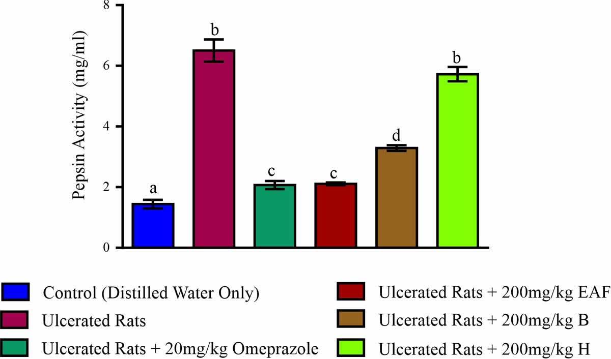
Conversely, ethylacetate fraction of A. mexicana leaf at 200 mg kg-1 b.wt., significantly attenuated the ulcer index (1.64±0.12 mm2), gastric volume (3.12±0.10 mL), gastric acidity (1.45±0.04 mL) and significantly increased the pH (4.54±0.16) to values comparable to the omeprazole treated group. The butanol fraction also showed a promising effect as it also attenuated the ulcer index (2.30±0.07 mm2), gastric volume (3.82±0.13 mL), gastric acidity (2.26±0.20 mL) and significantly (p<0.05) increased the pH values (3.97±0.08) when compared to the untreated group whereas the n-hexane fraction did not cause any significant alteration in the above ulcer indices when compared to the ulcerated group that do not receive medical intervention (Table 2). In Table 3, gastric H+/K+ ATPase activity of the ulcerated group (12.23±0.98 nmol-1 min-1 mg-1 protein) increased significantly (p<0.05) upon ulceration when compared to the control (7.68±0.37 nmol-1 min-1 mg-1 protein) however this proton pump activity was significantly (p<0.05) reduced in the ethylacetate fraction (7.74±0.47 nmol-1 min-1 mg-1 protein), butanol (8.32±0.20 nmol-1 min-1 mg-1 protein) fraction groups and reference drug (7.98±0.21 nmol-1 min-1 mg-1 protein) treated group when compared to the ulcerated untreated group. The ethylacetate fraction exhibited a better activity than the reference drug by significantly (p<0.05) reducing H+/K+ ATPase activity to value similar with the control (7.68±0.37 nmol-1 min-1 mg-1 protein). On the other hand, the activity of H+/K+ ATPase in the hexane fraction group (12.17 ± 1.06 nmol/min/mg protein) was not significantly (p > 0.05) different with the ulcerated group left with no medical intervention (12.23±0.98 nmol-1 min-1 mg-1 protein). Figure 1 depicts pepsin activity in the gastric juice of ulcerated rats treated with the three solvent-partitioned fractions of A. mexicana leaf. Administration of indomethacin significantly increased (p<0.05) the pepsin activity (6.4 mg mL-1) in the ulcerated untreated group when compared to the control (1.8 mg mL-1).
|
|||||
| Treatment Groups | ATPase Activity (nmol min-1 mg-1 protein) | ||||
| Control (Distilled Water) | 7.68 ± 0.37a | ||||
| Ulcerated Rats | 12.23 ± 0.98b | ||||
| Ulcerated Rats + 20 mg kg-1 Omeprazole | 7.98 ± 0.21a | ||||
| Ulcerated Rats + 200 mg kg-1 EAF | 7.74 ± 0.47a | ||||
| Ulcerated Rats + 200 mg kg-1 BF | 8.32 ± 0.20c | ||||
| Ulcerated Rats + 200 mg kg-1 HF | 12.17 ± 1.06b | ||||
Data are means of six determinations ± SEM. Values with different superscripts down the column for each parameter are significantly different (p<0.05) |
|||||
EAF: Ethylacetate Fraction, BF: Butanol Fraction, HF: Hexane Fraction |
|||||
 |
||||||||||||||||||||||||||||||||||||||||||||||||||||||||||||||||||||||||||||||||||||||||||||||||||||||||||||||||||||||||||||||||||||||||||||||||||||||||||||||||||||||||||||||
| |
| |
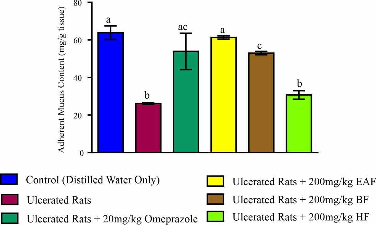
Treatment of ulcerated rats with each of 200 mg kg-1 b.wt., of ethylacetate and butanol fractions significantly (p<0.05) reduced the activity of pepsin to 2 and 3.2 mg mL-1 respectively when compared to the ulcerated group left untreated. Similarly, the ethylacetate fraction demonstrated the best efficacy as it was able to significantly (p<0.05) decrease pepsin activity in a manner comparable to the reference drug (2 mg mL-1). The adherent mucus content in ulcerated rats that received no medical intervention (25 mg/tissue) reduced significantly (p<0.05) when compared to the control (63 mg/tissue). However, the administration of 200 mg kg-1 b.wt., of ethylacetate fraction resulted to a significant increase (p<0.05) in the adherent mucus content (60 mg/tissue) in the stomach of rats and compete favorably well with the reference drug (56 mg/tissue) treated group (Fig. 2). Also the adherent mucus content of the butanol fraction treated (55 mg/tissue) rats was not significantly (p>0.05) different from the reference drug treated group (56 mg/tissue). Table 4 depicts the effect of administration of the solvent-partitioned fractions of A. mexicana leaf on the glycoprotein concentration in the stomach of indomethacin-induced ulcerated rats. The administration of indomethacin resulted to a significant depletion (p<0.05) in the total carbohydrate concentration (58.24±0.22 ug mL-1), glycoprotein concentration (1.07±0.21) and a significant increase (p<0.05) in the total protein concentration (54.38±0.21 ug mL-1) of ulcerated rats when compared to the control (130.36 ± 0.42, 3.05 ± 0.21 and 54.38 ± 0.16 ug mL-1 respectively). The glycoprotein concentration was significantly (p<0.05) increased upon treatment of rats with the three fractions when compared to the ulcerated group that received no medical intervention.
 |
|||||||||||||||||||||||||||||||||||||||||||||||||||||||||||||||||||||||||||||||||||||||||||||||||||||||||||||||||||||||||||||||||||||||||||||||||||||||||||||||||||||||||||
| |
| EAF: Ethylacetate Fraction, BF: Butanol Fraction, HF: Hexane Fraction |
|
|||||
| Treatment Groups | Total CHO (ug mL-1) | Total Protein (ug mL-1) | Total CHO:Total Protein (Glycoprotein) | ||
| Control (Distilled Water) | 130.36 ± 0.42a | 43.50 ± 0.12a | 3.05 ± 0.13a | ||
| Ulcerated Rats | 58.24 ± 0.20b | 54.38 ± 0.16b | 1.07± 0.21b | ||
| Ulcerated Rats + 20 mg kg-1 Omeprazole | 129.12 ± 0.21a | 42.31 ± 0.23a | 3.03 ± 0.11a | ||
| Ulcerated Rats + 200 mg kg-1 EAF | 130.10 ± 0.42a | 44.17 ± 0.03a | 3.08 ± 0.01a | ||
| Ulcerated Rats + 200 mg kg-1 BF | 117.86 ± 0.24c | 41.32 ± 0.01a | 2.54 ± 0.01c | ||
| Ulcerated Rats + 200 mg kg-1 HF | 54.94 ± 0.10b | 52.32 ± 0.24b | 1.12 ± 0.01b | ||
Data are means of six determinations ± SEM. Values with superscripts different from the control down each column for each parameter are significantly different (p<0.05) |
|||||
CHO: Carbohydrate, EAF: Ethylacetate Fraction, BF: Butanol Fraction, HF: Hexane Fraction |
|||||
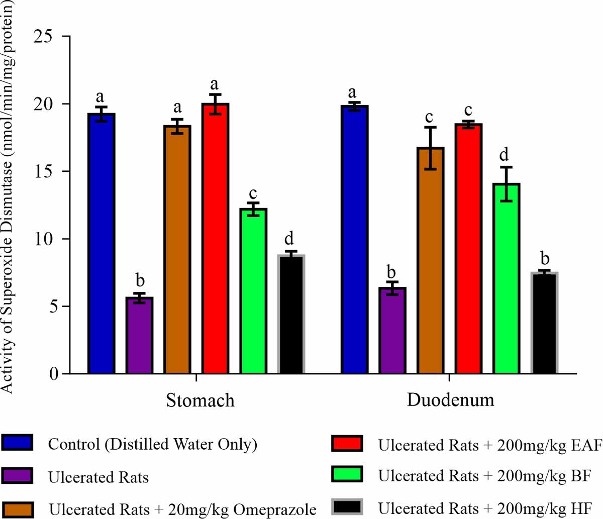
The glycoprotein concentration of rats that received the ethylacetate fraction (130.05±0.42) was similar to the concentration of glycoprotein observed in the omeprazole treated group (129.05±0.21) and the control (130.36±0.42). In addition, the glycoprotein concentrations in both the butanol fraction treated group (117.86±0.24) and the n-hexane treated group (54.94±0.10) were significantly lower when compared to the control and the omeprazole treated group. Figures 3 and 4 depict the activities of superoxide dismutase (SOD) and catalase (CAT) activities in the stomach and duodenum of ulcerated rats treated with solvent-partitioned fractions of A. mexicana leaf. Activities of superoxide dismutase and catalase were significantly (p<0.05) reduced in the stomach (5.50 nmol-1 min-1 mg-1 protein for SOD, 45.71 nmol-1 min-1 mg-1 protein for CAT) and duodenum (7.10 nmol-1 min-1 mg-1 protein for SOD, 25.43 nmol-1 min-1 mg-1 protein for CAT) of ulcerated rats with no medical treatment when compared to the control where the activities of SOD and CAT were 19.80 and 92.65 nmol-1 min-1 mg-1 protein in the stomach respectively and 20.02 nmol-1 min-1 mg-1 protein for SOD, 70.51 nmol-1 min-1 mg-1 protein for CAT in the duodenum of the control rats . Treatment with 200 mg kg-1 each of ethylacetate, butanol and n-hexane fractions significantly (p<0.05) upsurge the activity of SOD to 20.03, 13.24 and 9.48 nmol-1 min-1 mg-1 proteins in the stomach of rats respectively when compared to the ulcerated group left untreated whereas in the duodenum, only the ethylacetate fraction (18.02 nmol-1 min-1 mg-1 protein) and butanol fraction (14.76 nmol-1 min-1 mg-1 protein) significantly (p<0.05) elevated the activity of this enzyme (Fig. 3).
 |
||||||||||||||||||||||||||||||||||||||||||||||||||||||||||||||||||||||||||||||||||||||||||||||||||||||||||||||||||||||||||||||
| |
On a similar note, both ethylacetate and butanol fractions significantly (p<0.05) increased the activity of CAT in stomach (91.03 and 70.70 nmol-1 min-1 mg-1 protein respectively) and in the duodenum (72.24 and 70.53 nmol-1 min-1 mg-1 protein respectively) of ulcerated rats. The ethylacetate fraction increased the activities of SOD (20.03 and 18.02 nmol-1 min-1 mg-1 protein in the stomach and duodenum of the rats respectively) and CAT (91.03 and 72.24 nmol-1 min-1 mg-1 protein in the stomach and duodenum of the rats respectively) similar to the activities observed in the omeprazole treated group (19.60, 90.75 nmol-1 min-1 mg-1 protein for SOD and CAT in the stomach respectively and 17.40, 70.94 nmol-1 min-1 mg-1 protein in the duodenum respectively) (Fig. 3 and 4). There was depletion in the level of reduced glutathione (GSH) in the stomach (0.19 ± 0.01 nmol-1 mg-1 protein) and duodenum (0.17±0.01 nmol-1 mg-1 protein) of ulcerated rats when compared to the control (58.15±0.25 and 28.50±0.44 nmol mg-1 protein respectively). Upon treatment of animals with ethylacetate and butanol fractions, the level of glutathione significantly (p<0.05) increased in both tissues of rats when compared to the indomethacin-ulcerated rats as this fraction remarkably boosted the GSH level (0.41±0.01 nmol mg-1 protein in the stomach and 0.55±0.02 nmol mg-1 protein in the duodenum) better than the other two fractions (Table 5).
 |
||||||||||||||||||||||||||||||||||||||||||||||||||||||||||||||||||||||||||||||||||||||||||||||||||||||||||||||||||||||||||||
| |
| EAF: Ethylacetate Fraction, BF: Butanol Fraction, HF: Hexane Fraction |
|
||||||
| Treatment Groups | Reduced Glutathione (nmol mg-1 protein) | Glutathione Peroxidase (nmol mg-1 protein) | ||||
| Stomach | Duodenum | Stomach | Duodenum | |||
| Control (Distilled Water) | 0.52 ± 0.04a | 0.53 ± 0.03a | 58.15 ± 0.25a | 28.50 ± 0.44a | ||
| Ulcerated Rats | 0.19 ± 0.01b | 0.17 ± 0.01b | 31.28 ± 0.30b | 10.39 ± 0.09b | ||
| Ulcerated Rats + 20 mg kg-1 Omeprazole | 0.33 ± 0.03c | 0.55 ± 0.02ac | 57.62 ± 0.12a | 19.23 ± 0.04c | ||
| Ulcerated Rats + 200 mg kg-1 EAF | 0.41 ± 0.01ac | 0.48 ± 0.04c | 58.35 ± 0.37a | 19.49 ± 0.12c | ||
| Ulcerated Rats + 200 mg kg-1 BF | 0.31 ± 0.02c | 0.37 ± 0.03c | 56.80 ± 0.35a | 14.29 ± 0.12d | ||
| Ulcerated Rats + 200 mg kg-1 HF | 0.23 ± 0.02b | 0.24 ± 0.01d | 39.41 ± 2.11c | 12.23 ±0.10e | ||
Data are means of six determinations ± SEM. Values with different superscripts down column for each parameter are significantly different (p<0.05) |
||||||
EAF: Ethylacetate Fraction, BF: Butanol Fraction, HF: Hexane Fraction |
||||||
The 200 mg kg-1 b.wt., each of the ethylacetate, butanol and n-hexane fractions upon administration to animals significantly (p<0.05) elevated the activity of glutathione peroxidase which initially reduced in the stomach and duodenum of ulcerated rats left untreated. More so, the ethylacetate treated group elevated the activity of this enzyme (19.49±0.02 nmol mg-1 protein) in the stomach of rats to a manner that leveled up with the omeprazole treated group (19.23±0.04 nmol mg-1 protein). Table 6 depicts the activities of glutathione reductase and glutathione transferase in the stomach and duodenum of ulcerated rats treated with each of the fractions of A. mexicana leaf. A significant depletion (p<0.05) in the activity of glutathione reductase was observed in the stomach (31.28±0.30 nmol mg-1 protein) and duodenum (10.39±0.09 nmol mg-1 protein) of indomethacin-induced ulcerated rats when compared to the control (58.15±0.25 nmol mg-1 protein in stomach and 28.50±0.44 nmol mg-1 protein in the duodenum). This activity was however significantly (p<0.05) enhanced in both tissues of rats administered ethylacetate fraction (58.35±0.37 nmol mg-1 protein in stomach and 19.49±0.12 nmol mg-1 protein in duodenum) and butanol fraction (56.80±0.35 nmol mg-1 protein in the stomach and 14.29±0.17 nmol mg-1 protein). The activity of glutathione transferase significantly (p<0.05) increased in the duodenum of rats that received all fractions compared to the ulcerated group with no treatment whereas in the stomach of rats, only ethylaceatate and butanol fractions significantly elevated (p<0.05) the activity of glutathione transferase. The result showed a better activity of glutathione transferase in both tissues for the ethylacetate fraction and was comparable to the omeprazole treated group (Table 6).
|
||||||
| Treatment Groups | Glutathione Reductase (nmol mg-1 protein) | Glutathione-s-Transferase (nmol mg-1 protein) | ||||
| Stomach | Duodenum | Stomach | Duodenum | |||
| Control (Distilled Water) | 58.15 ± 0.25a | 28.50 ± 0.44a | 10.32 ± 0.92a | 2.10 ± 0.07a | ||
| Ulcerated Rats | 31.28 ± 0.30b | 10.39 ± 0.09b | 3.68 ± 0.14b | 0.87 ± 0.09b | ||
| Ulcerated Rats + 20 mg kg-1 Omeprazole | 57.62 ± 0.12a | 19.23 ± 0.04c | 9.32 ± 0.22c | 1.95 ± 0.03a | ||
| Ulcerated Rats + 200 mg kg-1 EAF | 58.35 ± 0.37a | 19.49 ± 0.12c | 9.63 ± 0.62c | 1.97 ± 0.04a | ||
| Ulcerated Rats + 200 mg kg-1 BF | 56.80 ± 0.35a | 14.29 ± 0.17d | 7.68 ± 0.42d | 1.83 ± 0.06c | ||
| Ulcerated Rats + 200 mg kg-1 HF | 39.41 ± 2.11b | 12.25 ± 0.30b | 3.73 ± 0.32b | 1.28 ± 0.06d | ||
Data are means of six determinations ± SEM. Values with different superscripts down each column for each parameter are significantly different (p<0.05) |
||||||
EAF: Ethylacetate Fraction, BF: Butanol Fraction, HF: Hexane Fraction |
||||||
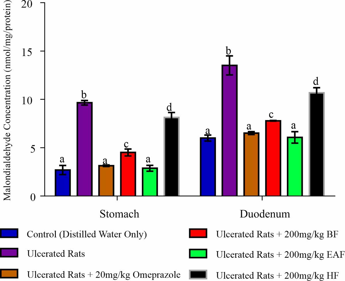
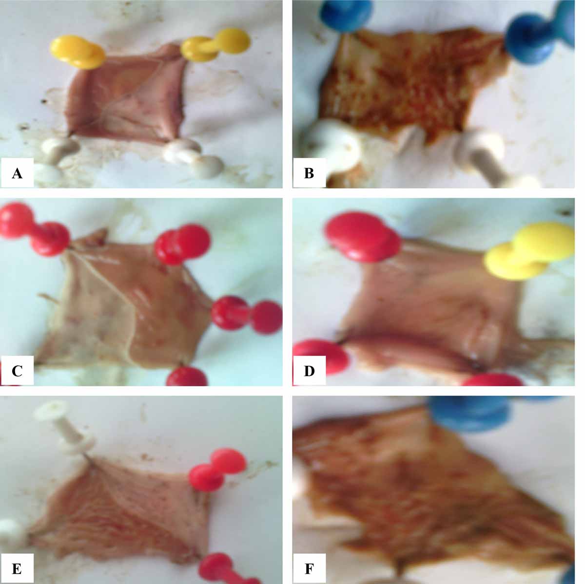
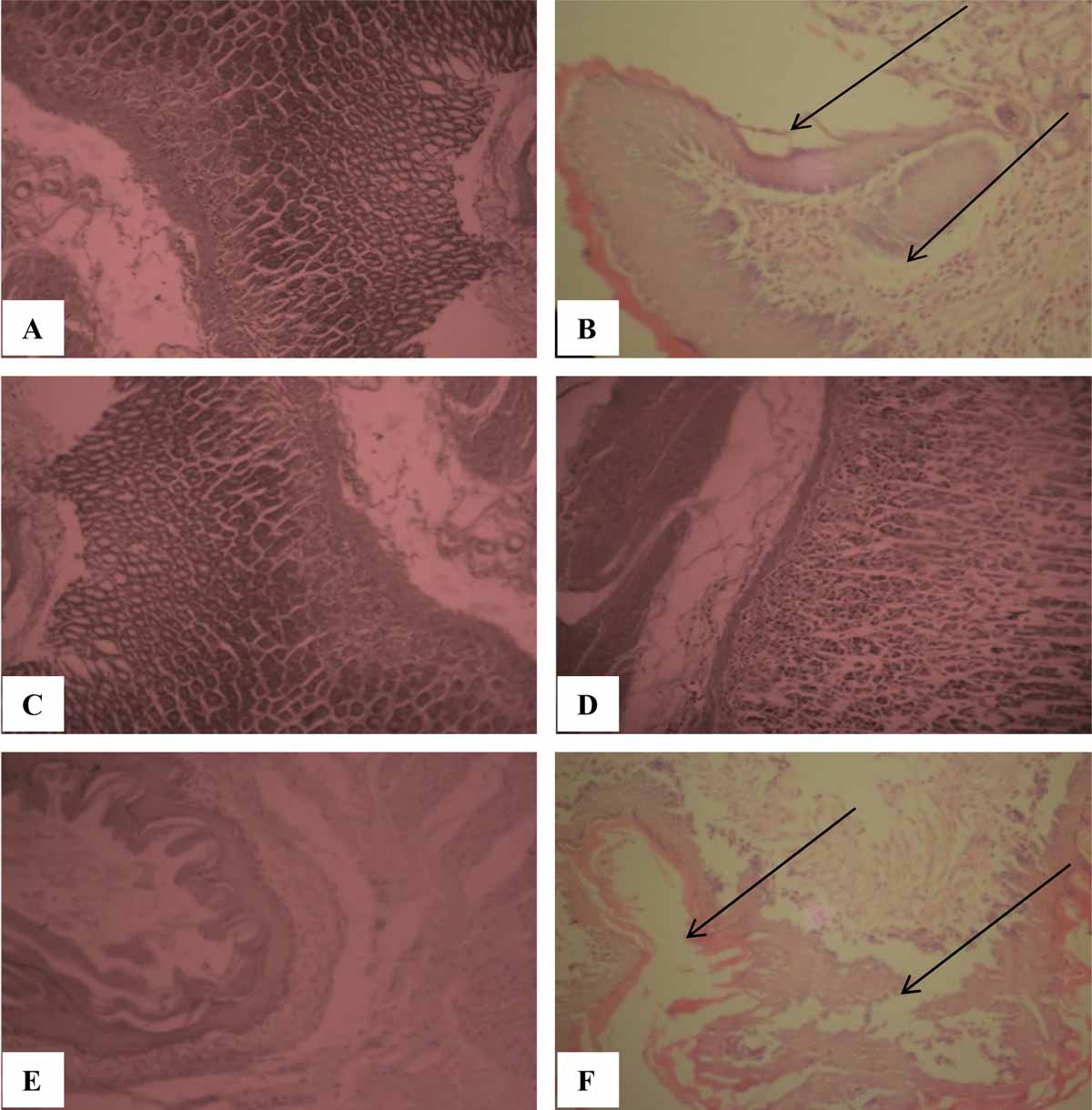
A significant upsurge (p<0.05) in malondialdehyde (MDA) concentration was observed in the stomach (9.85 mol mg-1 protein) and duodenum (13.95 mol mg-1 protein) of ulcerated rats when compared to the control (3.52 and 6.50 mol mg-1 protein respectively). MDA concentration significantly (p<0.05) reduced in both tissues of the animals treated with each of the fractions. Furthermore, the ethylacetate fraction recorded a better MDA reduction efficacy of 4.74 and 7.40 mol mg-1 proteins in the stomach and duodenum respectively than other two fractions (3.50, 6.35 mol-1 mg protein for butanol fraction and 8.45 mol mg-1 protein, 10.75 mol mg-1 protein for and hexane fraction in the stomach and duodenum respectively) when compared to the control and rats that received 20 mg kg-1 b.wt., omeprazole which recorded 3.75 and 6.90 mol mg-1 proteins reduction in MDA concentration in the stomach and duodenum respectively (Fig. 5). The macroscopic view of the stomach of the control rats showed no ulcer lesion (Fig. 6a) whereas the stomach of ulcerated animals that received no medical attention manifested ulcer lesion with severe hemorrhage (Fig. 6b). Rats treated with 20 mg kg-1 b.wt., omeprazole (Fig. 6c) and ethylacetate fraction (Fig. 6d) showed prominent amelioration of ulcer lesions in the stomach relative to the control (Fig. 6a). Treatment of rats with the butanol fraction also demonstrated promising gastric lesion amelioration (Fig. 6e) while ulcer lesion with appearance of hemorrhage was observed in the stomach of ulcerated rats that received the n-hexane fraction of A. mexicana leaf (Fig. 6f). Histopathological study on the control rats revealed a normal appearance and architecture of the gastric mucosal layers (Fig. 7a) whereas ulcerated rats with no medical intervention demonstrated a severe degeneration of gastric mucosal layers in the stomach tissue, (Fig. 7b) when compared to the control (Fig. 7a). Treatments of rats with omeprazole (Fig. 7c), ethylacetate (Figure 7d) and butanol fractions (Fig. 7e) of A. mexicana leaf ameliorated the degenerated layers of the gastric pit. Severe degeneration of the mucosal layer with a mild amelioration was noticed in rats that received n-hexane fraction as medication (Fig. f7).
 |
|||||||
| |
| EAF: Ethylacetate Fraction, BF: Butanol Fraction, HF: Hexane Fraction |
 |
||||
| |
| |
 |
|
| |
| |
DISCUSSION
The ethylacetate fraction as evident from this study showed the best anti-ulcerogenic activity. The activity possessed by this fraction maybe attributed to alkaloids and flavonoids which are the only secondary metabolites present in this fraction. Alkaloids and flavonoids are phytochemicals whose anti-ulcer activities have been established. Alkaloids such as berberine and ephedrine derived from the plant family Papaveraceae in which Argemone mexicana belongs are known to impair ulcer lesions produced by aggressive agents and alter the secretion of gastric acid by increasing the luminal gastric output of basal bicarbonate and pH28. Flavonoids have been reported to exhibit anti-ulcerogenic activity by inhibiting gastric acid secretion and offer gastroprotection by increasing gastric blood flow, stimulating mucus content and increasing the protective effects of prostaglandins on the gastric mucosal29. Indomethacin a Non-steroidal anti-inflammatory drug (NSAID) used to induce ulcer in this study is one of the ulcer models that has gained acceptance in assessing the anti-ulcerogenic activity of a significant number of plants.
The increase in gastric acidity of ulcerated rats maybe due to the suppression of prostaglandin by indomethacin which act through inhibition of cyclo-oxygenase, an enzyme responsible for the biosynthesis of prostaglandin that stimulate the secretion of other defensive factors (mucus, glycoprotein) and subsequently increasing stomach gastric output. The ulcer index measures the level of excavation or lesion on the gastric mucosal. The increase in ulcer index of ulcerated rats is an indication of assault on the gastric mucosal which may have resulted from the imbalance between the acid/pepsin and defensive factors of the mucosal caused by indomethacin. It has been reported that NSAIDs like indomethacin are capable of causing an imbalance between gastric offensive factor and defensive factors2.
The pH is a reflection of gastric acidity known to accompany increased gastric acid secretion. Therefore it is evident from this study that increased gastric acidity resulted to the decrease in pH value observed in the ulcerated group that received no medical intervention. The increase in gastric acid volume is also a reflection of increased gastric acid secretion as gastric acid secretion is a determinant of the gastric acid volume and increased gastric volume is known to correlate to increased acid secretion. The attenuation of these ulcerative indices by the ethylacetate and butanol fractions may indicate that the fractions established equilibrium between the offensive and defensive factors due to the presence of flavonoids which stimulates prostaglandin synthesis. Flavonoids have been reported to increase prostaglandin production30 thus subsequently enhance the mucosal barrier against the damaging effect of acid.
The proton pump also called the H+/K+ ATPase enzyme is the terminal stage involve in gastric acid secretion and it is being directly responsible for the secretion of H+ into the lumen of the gastric mucosal in exchange for K+ using the energy derived from the hydrolysis of ATP. The increase in the H+/K+ ATPase enzyme may indicate that indomethacin activates or cause the up-regulation of the proton pump thereby subsequently increasing the gastric output in the stomach. The decrease in the activity of this pump which was more remarkable in the ethylacetate treated rats may be attributed to the presence of flavonoids causing the down-regulation of the pump. Zerumbone, gingerol and zingerone are flavonoids isolated from Zingiber officinalis and have shown significant influence in inhibiting parietal H+/K+ ATPase30. Pepsin is responsible for proteolysis in the stomach and its activity depends on gastric acid output since gastric acid is required for its acidification from pepsinogen to pepsin. The increase in pepsin activity of ulcerated rats may have resulted from the activated proton pump which increased the gastric output and subsequently increasing the pepsin activity since gastric acid is required for acidification of this proteolytic enzyme. Both mucus and glycoprotein constitute the gel layer that coat and promote the thickness of the gastric mucosal barrier. The increase in adherent mucus content and glycoprotein concentration in ulcerated rats administered 200 mg kg-1 b.wt., of the ethylacetate and butanol fractions which decreased in ulcerated rats that received no medical intervention indicates that both fractions were able to strengthen the mucosal layer preventing it from the corrosive effect of acid and pepsin. Cellular antioxidant enzymes such as superoxide dismutase (SOD), catalase (CAT), glutathione reductase (GSH-Red), glutathione peroxidase (GSH-Px), glutathione-S-transferase (GST) and non-enzymic antioxidant reduced glutathione (GSH) act as the first line of defense against oxidative injury31 and once damaged ulceration may result. The decrease in activities of SOD, CAT, GSH-Red, GSH-Px and GST in ulcerated rats is an indication of increased oxidative stress on the gastric mucosa of rats induced by free radicals resulting to an imbalance between peroxidant and antioxidant enzymes.
Oxidative stress has been implicated in the pathogenesis of indomethacin-mediated ulceration32. The increase in the activities of these antioxidant enzymes in the stomach and duodenum of rats following their treatment with the ethylacetate and butanol fractions indicates that the fraction restored normalcy from the assault caused by free radicals on these antioxidant enzymes. This boost in the antioxidant status may have been facilitated by flavonoids because flavonoids are known to play significant role in the stabilization of the antioxidant enzymes33. Superoxide dismutase is a metal complexed enzyme that dismutase superoxide anion to hydrogen peroxide and oxygen. Hydrogen peroxide formed from dismutation reaction catalyzed by superoxide dismutase is capable of penetrating the membrane where it decomposes into hydroxyl radical a more powerful and damaging radical therefore, scavenging hydrogen peroxide is necessary. Catalase and glutathione peroxidase function as enzymes to scavenge hydrogen peroxide by reducing it to water as well as water and oxygen respectively. GSH-Px also catalyzes the hydrolysis of hydroperoxides to their corresponding alcohols by using reduced glutathione (GSH) which is later oxidized to glutathione disulfide (GSSG). Glutathione reductase (GSH-Red) generates and recycles GSH for GSH-Px as substrate to scavenge radicals and reduce hydrogen peroxide. The depleted level of GSH may also be due to the reduction in the activity of GSH-Red resulting in concomitant reduction in the level of GSH since GSH-Red is known to recycle GSH. The level of glutathione which remarkably increased in ulcerated rats treated with ethylacetate and fractions in this study might have resulted from the increased activity of GSH-Red which replenishes GSH by causing reducing GSSG. Lipid peroxidation is the oxidative deterioration of polyunsaturated fatty acids of the lipid components of the plasma membrane.
Lipid peroxidation result from the reaction of radicals such as hydroxyl radical with lipid component of the plasma membrane and malondialdehyde is a product used as a major biomarker for lipid peroxidation. The increased concentration of malondialdehyde observed in ulcerated rats is a reflection of increased oxidative stress induced on the gastric mucosal membrane induced by indomethacin which is one of the mechanisms of indomethacin in ulcer formation34,35. The reversal of the lipid peroxidation in rats treated with the fractions (particularly ethylacetate and butanol) is an indication that the fractions caused a marked suppression in oxidative stress facilitated by the presence of flavonoids in both fractions acting as free radical scavengers in lipid peroxidation31.
Histological changes are late manifestation of assault on tissues36. The severe degenerated architecture of the stomach is an indication of level of assault caused by indomethacin on sub-mucosal layer of the stomach as seen in the histological and microscopic view of the stomach. Treatment of rats with ethylacetate fraction of A. mexicana administered at 200 mg kg-1 was able to ameliorate the assault caused by indomethacin on the gastric mucosa and this amelioration was similar to what was observed in the omeprazole group. This is an indication of the wound healing property of the plant facilitated by the presence of flavonoids. From the study, the ethylacetate fraction exhibited better activity in attenuating the acid-secretory parameters and as well ameliorating the antioxidant status of ulcerated rats. Similarly, the butanol fraction also exhibited a promising effect thus the study suggest that the bioactive type of flavonoids responsible for the anti-ulcerogenic activity of A. mexicana may actually resides in the ethylacetate fraction than the butanol fraction since the ethylacetate fraction was the most effective. In view of this, there is need to further isolate the flavonoids in order to establish this.
CONCLUSION
The study validates the anti-ulcerogenic activity of Argemone mexicana leaf as acclaimed in folk medicine of Nigeria with evidence of attenuation of acid secretory parameters, enhancement of both mucosa mucus content and antioxidant enzymes in the stomach and duodenum of rats. Study is already on going to isolate and identify the active principles in this plant and to propose their mechanism of action.
SIGNIFICANCE STATEMENT
This study discovered that partitioned fractions of Argemone mexicana leaves can offer gastro-protection and ameliorate gastric lesions in rats which can be beneficial for treating peptic ulcer. This study will help in the search for the development of a more effective anti-ulcerogenic drug of plant origin.
REFERENCES
- Prasad, B., A. Wei and B. D, 2012. Pharmacology of traditional herbal medicines and their active principles used in the treatment of peptic ulcer, diarrhoea and inflammatory bowel disease. New Adv. Basic Clin. Gastroenterol.
- Søreide, K., K. Thorsen, E.M. Harrison, J. Bingener, M.H. Møller, M. Ohene-Yeboah and J.A. Søreide, 2015. Perforated peptic ulcer. Lancet, 386: 1288-1298.
- Levenstein, S., S. Rosenstock, R.K. Jacobsen and T. Jorgensen, 2015. Psychological stress increases risk for peptic ulcer, regardless of Helicobacter pylori infection or use of nonsteroidal anti-inflammatory drugs. Clin. Gastroenterol. Hepatol., 13: 498-506.e1.
- Das, A.K., P. Bigoniya, N.K. Verma and A. Rana, 2012. Gastroprotective effect of Achyranthes aspera linn. leaf on rats. Asian Pac. J. Trop. Med., 5: 197-201.
- De Sousa Falcao, H., J.A. Leite, J.M. Barbosa-Filho, P.F. de Athayde-Filho and M.C. de Oliveira Chaves et al., 2008. Gastric and duodenal antiulcer activity of alkaloids: A review. Molecules, 13: 3198-3223.
- Magaji, R.A., M.A.M. Okasha, M.S. Abubakar and M.Y. Fatihu, 2007. Anti-ulcerogenic and Anti-secretory activity of the N-butanol portion of Syzygium aromaticum in Rat. Niger. J. Pharm. Sci., 6: 119-126.
- Siddiqui, I.A., S.S. Shaukat, G.H. Khan and M.J. Zaki, 2002. Evaluation of Argemone mexicana for control of root-infecting fungi in potato. J. Phytopathol., 150: 321-329.
- Brahmachari, G., D. Gorai and R. Roy, 2013. Argemone mexicana: Chemical and pharmacological aspects. Rev. Bras. Farmacogn., 23: 559-575.
- Sourabie, T.S., H.M. Kone, J.B. Nikiema, O.G. Nacoulma and I.P. Guissou, 2010. Evaluation of the antihepatotoxic effect of Argemone Mexicana leaf extracts against ccl4-induced hepatic injury in rats. Int. J. Bio. Chem. Sci., 3: 1499-1503.
- Harborne, J.B., 2011. Phytochemical methods: a guide to modern techniques of plant analysis. 1st Edn., Springer, Cham, Amsterdam, Netherlands.
- Sofowora, A., 1996. Research on medicinal plants and traditional medicine in Africa. J. Alt. Compl. Med., 2: 365-372.
- Trease, G.E. and W.C. Evans, 1989. A Textbook of Pharmacognosy. 13th Edn., Bailliere Tindall Ltd., London.
- Marslin, G., B. Divya, R.A. Mary, M.M.H. Viji, V.K. Kalaichelvan and V. Palanivel, 2013. Anti–ulcer activity of Ficus religiosa leaf ethanolic extract. Asian Pac. J. Trop. Biomed., 3: 554-556
- Maity, S., J.R. Vedasiromoni and D.K. Ganguly, 1995. Anti-ulcer effect of the hot water extract of black tea (Camellia sinensis). J. Ethnopharmacol., 46: 167-174
- Hirohashi, M., K. Takasuna, Y. Kasi, C. Usui, K. Tamura and H. Kojima, 1993. General pharmacological profile of the new anti-ulcer drug 3-[[[2-(3,4-dimethoxyphenyl)ethyl]carbamoyl]methyl]-amino-N-methylbenzamide. Arzneimittel-Forschung, 43: 569-577
- Nair, R.B. and P.A. Kurup, 1975. Investigations on the venom of the South Indian scorpion Heterometrus scaber. Biochim. Biophys. Acta (BBA)-Gen. Subj., 381: 165-174
- Lowry, O., N. Rosebrough, A.L. Farr and R. Randall, 1951. Protein measurement with the folin phenol reagent. J. Bio. Chem., 193: 265-275
- Reyes-Chilpa, R., C.H. Baggio, D. Alavez-Solano, E. Estrada-Muniz, F.C. Kauffman, R.I. Sanchez and S. Mesia-Vela, 2006. Inhibition of gastric H+, K+-ATPase activity by flavonoids, coumarins and xanthones isolated from Mexican medicinal plants. J. Ethnopharmacol., 105: 167-172
- AlRashdi, A.S., S.M. Salama, S.S. Alkiyumi, M.A. Abdulla, A.H.A. Hadi et al., 2012. Mechanisms of gastroprotective effects of ethanolic leaf extract of Jasminum sambac against HCl/Ethanol-induced gastric mucosal injury in rats. Evidence-Based Complement. Altern. Med.
- Ellman, G.L., 1959. Tissue sulfhydryl groups. Arch. Biochem. Biophys., 82: 70-77.
- Sun, M. and S. Zigman, 1978. An improved spectrophotometric assay for superoxide dismutase based on epinephrine autoxidation. Anal. Biochem., 90: 81-89.
- Beers, R.F. and I.W. Sizer, 1952. A spectrophotmeteric method for measuring breakdown of hydrogen peroxide by catalase. J. Bio. Chem.,, 195: 133-140.
- Faizal, P., B. Satheesan, B. Vinod and K.T. Augusti, 2017. Evaluation of antioxidant, lipid peroxidation and toxic effects after pomegranate intake in healthy human volunteers. Int. J. Clin. Med., 08: 12-20.
- Paglia, D.E. and W.N. Valentine, 1967. Studies on the quantitative and qualitative characterization of erythrocyte glutathione peroxidase. J. Lab. Clin. Med., 70: 158-169.
- Habig, W.H., M.J. Pabst and W.B. Jakoby, 1974. Glutathione S-transferases: The first enzymatic step in mercapturic acid formation. J. Biol. Chem., 249: 7130-7139.
- Buege, J.A. and S.D. Aust, 1978. Microsomal Lipid, Peroxidation. In: Methods in Enzymology, Vol. 52, Flesicher, S. and L. Packer (Eds.)., Academic Press, New York pp: 302-310.
- Krause, W.J., 2004. The Art of Examining and Interpreting Histologic Preparations: A Laboratory Manual and Study Guide for Histology. Universal-Publishers, Boca Raton, Florida, USA pp: 9-10.
- Yong, D.G., B.Q. Geng, G.G. Gu, F.M. Zhong and W.H. Yu, 1991. Anti-ulcer effect of anisodamine in rats. Acta Pharmacol., 12: 522-525.
- Awaad, A.S., R.M. El-Meligy and G.A. Soliman, 2013. Natural products in treatment of ulcerative colitis and peptic ulcer. J. Saudi Chem. Soc., 17: 101-124.
- Siddaraju, M.N. and S.M. Dharmesh, 2007. Inhibition of gastric H+, K+-atpase andhelicobacter pylori growth by phenolic antioxidants of Zingiber officinale. Mol. Nutr. Food Res., 51: 324-332.
- Abdallah, I.Z.A., H.A.H. Khattaba and G.H. Heeba, 2011. Gastroprotective effect of assyrian plum (Cordia myxa L.) fruit extract against indomethacin-induced gastric ulceration in rats. Life Sci. J.., 8: 433-445.
- Utsumi, H., K. Yasukawa, T. Soeda, K.I. Yamada, R. Shigemi, T. Yao and M. Tsuneyoshi, 2005. Noninvasive mapping of reactive oxygen species by in vivo electron spin resonance spectroscopy in indomethacin-induced gastric ulcers in rats. J. Pharmacol. Exp. Ther., 317: 228-235.
- Benchikh, F., 2018. Pharmacological effects of Myrtus Communis L. on the gastrointestinal tract of rats and mice. Ph.D. Thesis, Department of Biology and Animal Physiology, Faculty of Nature and Life Sciences. Ferhat Abbas Sétif 1 University, Setif, Algeria.
- Suleyman, H., A. Albayrak, M. Bilici, E. Cadirci and Z. Halici, 2010. Different mechanisms in formation and prevention of indomethacin-induced gastric ulcers. Inflammation, 33: 224-234.
- Wang, T., S. Zhao, Y. Wang, Y. Yang and L. Yao et al., 2014. Protective effects of escin against indomethacin-induced gastric ulcer in mice. Toxicol. Mech. Methods, 24: 560-566.
- Adesokan, A.A., M.A. Akanji and M.T. Yakubu, 2007. Antibacterial potentials of aqueous extract of Enantia chlorantha stem bark. Afr. J. Biotechnol., 6: 2502-2505.
How to Cite this paper?
APA-7 Style
Idowu,
O., Saliu,
O., Fakorede,
N., Arise,
R. (2021). Anti-Ulcerogenic Efficacy of Leaf Fractions of Argemone mexicana Against Indomethacin Induced-Ulceration in Rats. Asian Journal of Emerging Research, 3(2), 129-136. https://doi.org/10.3923/ajerpk.2021.129.136
ACS Style
Idowu,
O.; Saliu,
O.; Fakorede,
N.; Arise,
R. Anti-Ulcerogenic Efficacy of Leaf Fractions of Argemone mexicana Against Indomethacin Induced-Ulceration in Rats. Asian J. Emerg. Res 2021, 3, 129-136. https://doi.org/10.3923/ajerpk.2021.129.136
AMA Style
Idowu
O, Saliu
O, Fakorede
N, Arise
R. Anti-Ulcerogenic Efficacy of Leaf Fractions of Argemone mexicana Against Indomethacin Induced-Ulceration in Rats. Asian Journal of Emerging Research. 2021; 3(2): 129-136. https://doi.org/10.3923/ajerpk.2021.129.136
Chicago/Turabian Style
Idowu, O.A., O.A. Saliu, N.C. Fakorede, and R.O. Arise.
2021. "Anti-Ulcerogenic Efficacy of Leaf Fractions of Argemone mexicana Against Indomethacin Induced-Ulceration in Rats" Asian Journal of Emerging Research 3, no. 2: 129-136. https://doi.org/10.3923/ajerpk.2021.129.136

This work is licensed under a Creative Commons Attribution 4.0 International License.
Division of Scientific Publishing
ACE College for Women
Faisalabad-38090, Pakistan
ISSN: 2663-4988 / 2664-5211
This work is licensed under a Creative Commons Attribution 4.0 International License.