
Orally Administered Bisphenol A Impairs Normal Thyroid Functions Altering Thyroid Hormones Homeostasis in Female Wistar Rats
-
Chinenye E. Oguazu


Department of Applied Biochemistry, Faculty of Biosciences, Federal University Otuoke, Bayelsa, Nigeria
Francis C. EzeonuDepartment of Applied Biochemistry, Faculty of Biosciences, Federal University Otuoke, Bayelsa, Nigeria
Ikimi G. CharlesDepartment of Applied Biochemistry, Faculty of Biosciences, Enugu State of Science and Technology, Enugu, Nigeria
Ani N. OnuabuchiDepartment of Applied Biochemistry, Faculty of Biosciences, Enugu State of Science and Technology, Enugu, Nigeria
Dike Dike C. CharlesDepartment of Human Biochemistry, College of Basic Health Sciences, Nnamdi Azikiwe University, Nnewi, Nigeria
| Received 16 Feb, 2021 |
Accepted 12 Apr, 2021 |
Published 15 Jun, 2021 |
Background Objective: Bisphenol A (BPA) is an endocrine-disrupting chemical used on a wide range in the industry. Several studies reported that BPA may cause cardiovascular disorders in humans and animals. The present study aims to investigate the effect of BPA on the thyroid gland of female rats. Materials and Methods: The rats received a daily oral administration of BPA (0.05 – 1.0 mg kg-1 for 13 weeks). Thyroid Stimulating Hormone (TSH), Thyroxine (T4) and Triiodothyronine (T3) were assayed using an Auto chemical analyzer and data obtained were subjected to statistical analysis with the Statistical Package for the Social Sciences (SPSS) software. Results: It was found that BPA at the eleven studied doses induced a significant increase in the Thyroxin Stimulating Hormone (TSH), Free Thyroxin (FT4), Total Thyroxin (TT4) level, while the bound thyroxin is low compared to the control. The Free Triiodothyronine (FT3) and Total Triiodothyronine (TT3) were initially low at group 1 but at the other doses, they were on the increase. The bound triiodothyronine is lower when compared to that of the control throughout the studied time intervals. Conclusion: These results suggest that BPA has thyroid toxic effects which are mediated by the oxidative stress resulting from the overproduction of free radicals, BPA may also participate in the thyroid gland function disturbances.
INTRODUCTION
Bisphenol-A is an organic compound that has two phenol functional groups. It is prepared by the condensation reaction of acetone, and two equivalents of phenol1 with hydrochloric acid2 (Fig. 1). Bisphenol-A has a phenolic odour witha melting point of 155°C and a specific gravity of 1.060-1.195 gcm-13,4. It is soluble in nonpolar solvent5. Aerobic biodegradation and biodegradation half-life for BPA in river water and soil is about 4.5 days3 and the photo-oxidation half-life for BPA in the air is about 4 hrs6. It is known that the global population is subject to repeated exposure to BPA is via the diet7 with detectable metabolites in the urine8. Daily dietary intake of about 0.2 μg kg-1 body weight in breastfed babies and 1.5 μg kg-1 body weight in adults9 relevant doses of BPA causes increases in weight and size of the prostate gland, decreases in sperm efficiency, earlier puberty10, and abnormalities in the oocytes11. Invernizzi12 showed that BPA triggers ductal and alveolar structures proliferations, development of ductal hyperplasia13, modifications of the mammary gland architecture14, mammary carcinogenesis15, inflammatory cytokine dysregulation16, and mitochondrial-mediated apoptosis17. Health implications associated with BPA exposure include diabetes18, cardiovascular disease19, altered liver enzymes activities20 and obesity-promoting effects21. BPA alters glucose homeostasis, increased pancreatic insulin and induced insulin resistance22, BPA induces oxidative stress23, coronary artery disease24, activates Maxi-K ion channels25, increased BP and decreased Heart Rate (HR)26, increased risk of hypertension27, decreased efficiency of sperm production23 and increased ovarian cancer cell proliferation28. According to this, Miyagawa et al.29 reported impaired memory, increased aggressiveness30, alters anxiety31, loss or reduction of sexual dimorphisms32, loss of sex difference in corticotrophin-releasing hormones33, reduced the amount of tyrosine by dioxylase-immunoreactive34. This study aims to establish the possible effects and physiological disposition of Bisphenol-A on sex and thyroid hormones, in female Wistar albino rats.
(CH3)2CO+2C6H5OH (CH3)2C(C6H4OH)2+H2O
 |
||||||||||||||
| |
MATERIALS AND METHODS
Study area: The study was carried out at Applied Biochemistry Lab, Nnamdi Azikiwe University, Awka, Nigeria and Biochemistry Lab, Gregory University Uturu, AbiaState, Nigeria from June-September, 2017.
Methodology: Total 60 non-pregnant female rats of 5 weeks age were acclimatized in the laboratory for 7 days and randomly divided into 11 experimental groups of 5 rats each and respectively administered; 0.05, 0.1, 0.2, 0.3, 0.4, 0.5, 0.6, 0.7, 0.8, 0.9 and 1 mg of BPA kg-1 b.wt.day-1. The first group which served as control did not receive any treatment, but distilled water instead. The graded doses of BPA were dissolved in distilled water and administered by oral gavage using an intubation cannula (Lars Medicare Pvt. Ltd, New Delhi, India). Blood was obtained from the tail of the various groups by capillary action, weekly, after BPA administration for 13 weeks. Blood samples were processed for clinical assay.
Animals have housed in aluminium wire-mesh cages in a well-ventilated animal house with a 12 hrs dark/light cycle and at room temperature and were provided commercial rat pellets (Vital feed from Vital group of Company, Nigeria) and water ad libitum.
At the end of the experiments serum TSH, Thyroxine (T4) and Triiodothyronine (T3) were assayed using an Auto chemical analyzer (Lx20 pro-Autoanalyzer, Beckman Coulter, Woerden, Netherland and Chemwell chemical Analyzer, Manufacturer: Roche Hitachi, GMI.). All reagents were commercially obtained as already prepared kits. The kits for TSH, Thyroxine (T4) and Triiodothyronine (T3) were purchased from Phoenix Pharmaceuticals, Burlingame, CA; Enzo Life Sciences Inc, Boulevard Farmingdale, NY; Diagnostic automation/Cortez Diagnostics Inc, Calabasas, CA. Individual tests were carried out according to the kit specifications.
Statistical analysis: Differences between obtained values (mean±SD) were carried out by one-way analysis of variance (ANOVA) using SPSS software version 20.0 followed by the Tukey-Kramer multiple comparison test. At p≤0.05 was taken as a criterion for a statistically significant difference.
RESULTS
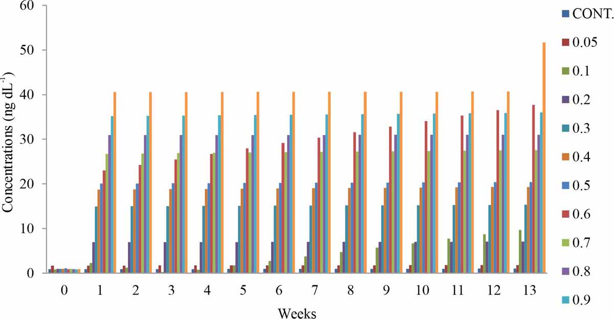
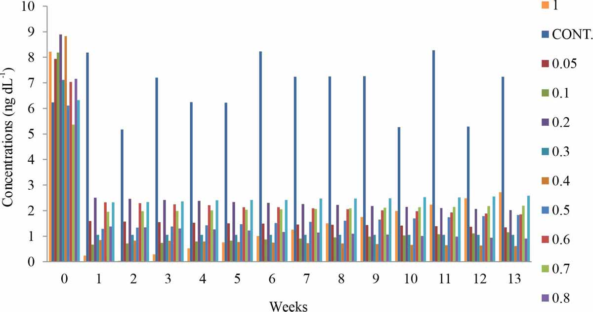
Thyroid-stimulating hormone: The experimental groups that were administered with 0.2–1 mg kg-1 BPA showeda dose-dependent significant increase in the concentration of thyroid-stimulating hormones when compared with the control (Fig. 2), the increase over time appeared to be relatively constant except for 0.6 mg kg-1 BPA test group that showed a constant increase in TSH with time and 1 mg kg-1 BPA test group that showed high levels of the thyroid-stimulating hormone at week 13. The test exposed 0.05 mg kg-1 BPA showed a nonsignificant increase in TSH that remained relatively constant with time. The group that received 0.1 mg kg-1 BPA responded differently during the study, it was observed that at the first week of exposure there was an increase in TSH, which gradually decreased through week 3, but surprisingly a consistent rise in the concentration of TSH recorded from week 4-13. All the weeks of exposure showed a characteristic dose-dependent effect of BPA on TSH, as the administered dose increases, the effect of BPA on TSH increases. The weeks of exposure showed sensitivity at two-point (0.1 and 0.6) but was constant at other points of exposure.
 |
||||||||||||
| |
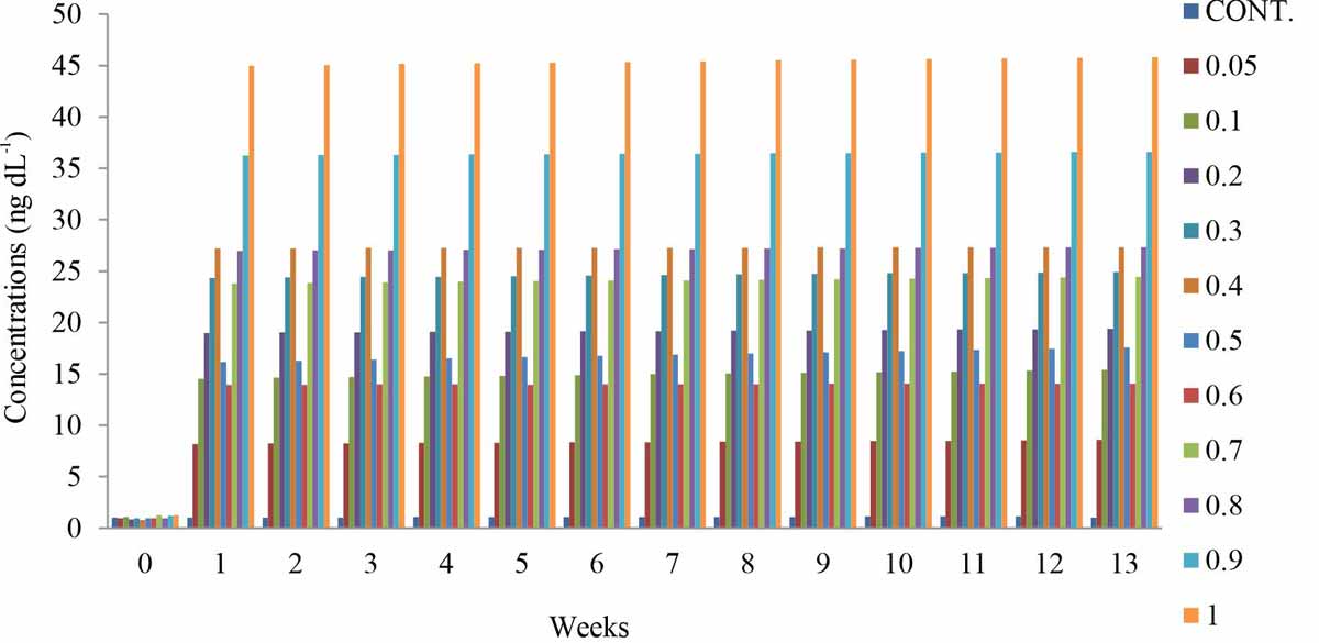
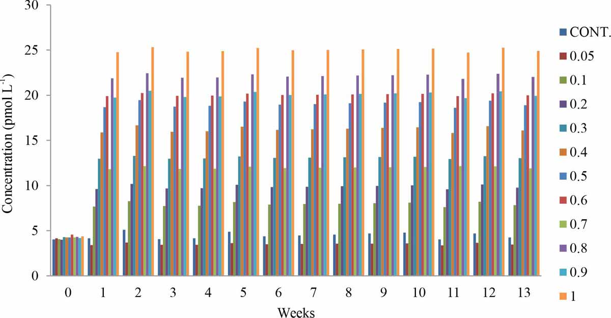
Total Thyroxine (TT4): All the experimental groups that were administered with (0.05–1 mg kg-1) BPA showed a significant increase in the concentration of TT4 when compared with the control (Fig. 3), amidst the increase TT4, the test groups exposed to 0.05-0.4 and 0.6-1 mg kg-1 showed a dose-dependent effect, while the TT4 level of the test groups exposed to 0.5 mg kg-1 BPA decreased relative to those of 0.4 mg kg-1 again those of 0.6 mg kg-1 test group decreased further relative to those of 0.5 mg kg-1 BPA test group. All the weeks of exposure showed a characteristic high but not dose-dependent effect of BPA on TT4. The weeks of exposure showed a non-sensitivity response with the lest observed effect at point 0.9.
 |
||||||||||
| |
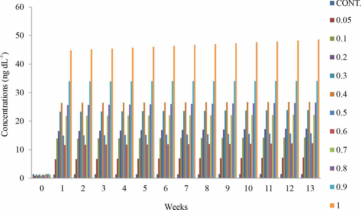
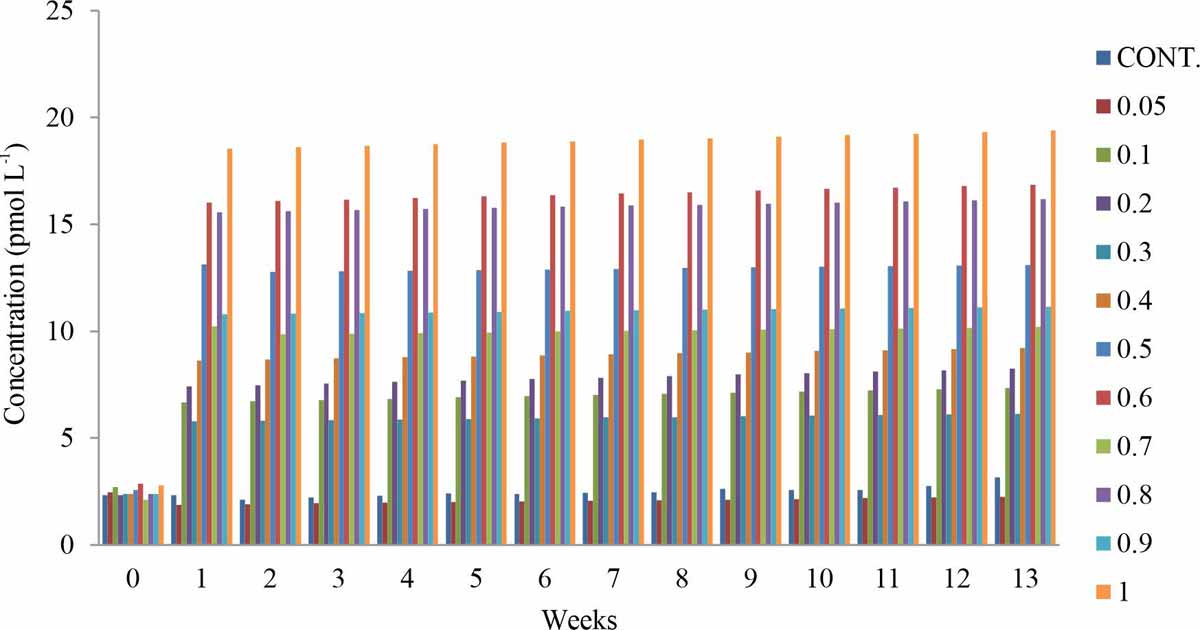
Free Thyroxine (FT4): All the experimental groups that were administered with (0.05–1 mg kg-1) BPA showed a significant increase in the concentration of FT4 when compared with the control (Fig. 4), which appears to be relatively constant with the time of exposure. Amidst the increase FT4, the test groups exposed to 0.05-0.4, and 0.6-1 mg kg-1 showed a dose-dependent effect, while the FT4 level of the test groups exposed to 0.5 mg kg-1BPA decreased relative to those of 0.4 mg kg-1 again those of 0.6 mg kg-1 test group decreased further relative to those of 0.5 mg kg-1 BPA test group. All the weeks of exposure showed a characteristic high and dose-dependent effect of BPA on FT4 on the 0.05–0.4 doses of BPA. The weeks of exposure showed a decrease response at point 0.5 -0.6 mg kg-1 BPA and tend to rise from 0.7 1 mg kg-1.
 |
||||||||
| |
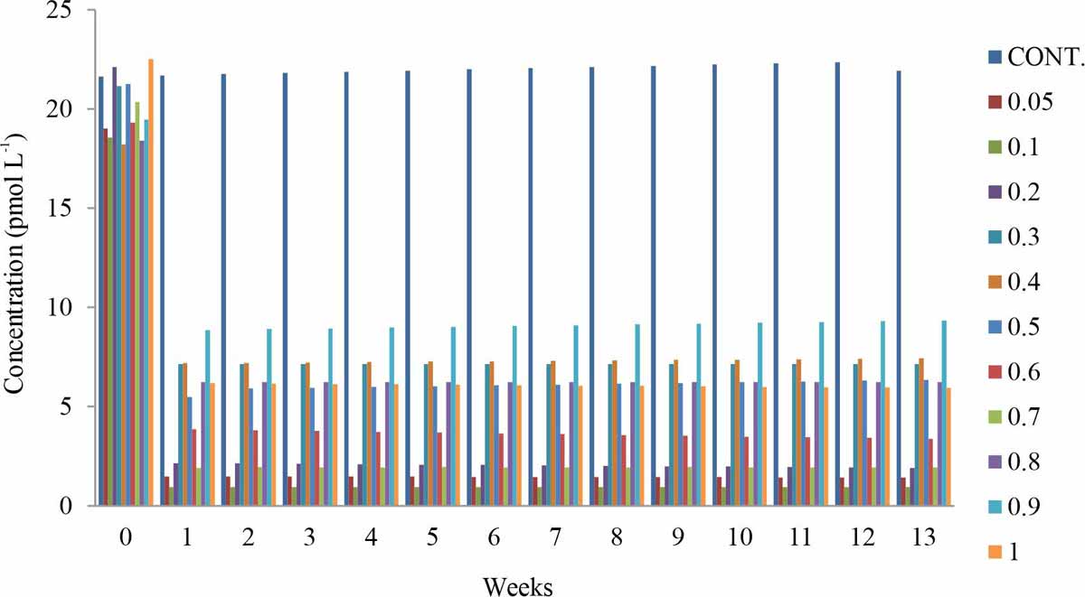
Conjugated Thyroxine (BT4): All the experimental groups that were administered with (0.05–1 mg kg-1) BPA showed a significant noncharacteristic decrease in the concentration of conjugated thyroxine (BT4) when compared with the control (Fig. 5). All the weeks of exposure showed a noncharacteristic low but not the dose-dependent effect of BPA on BT4. The weeks of exposure showeda slight sensitivity response with the highest observed effect at point 1 mg kg-1.
 |
||||||
| |
Total Triiodothyronine (TT3): The experimental groups that were administered with 0.1–1 mg kg-1 BPA showed a dose-dependent significant increase in the concentration of Total Triiodothyronine (TT3) when compared with the control (Fig. 6), the increase over time appeared to be relatively constant except 0.7 and 0.9 mg kg-1 BPA test group that showed a decrease in TT3 relative to 0.6 and 0.8 mg kg-1 BPA test group respectively. The test exposed 0.05 mg kg-1 BPA showed a nonsignificant decrease in TT3 that remained relatively constant with time (Fig. 6). All the weeks of exposure showed a characteristic relative dose-dependent effect of BPA on TT3, as the administered dose increases, the effect of BPA on TT3 increases. The weeks of exposure showed a low level of TT3 at 0.7 points, falling below the control at point 0.05.
 |
||||
| |
Free Triiodothyronine (FT3): The experimental groups that were administered with 0.1–1mg kg-1BPA showed a non-dose-dependent significant increase in the concentration of Free Triiodothyronine (FT3) when compared with the control (Fig. 7). The test exposed 0.05 mg kg-1 BPA showed a nonsignificant decrease in FT3 that remained relatively constant with time (Fig. 7). All the weeks of exposure showed a characteristic relative effect of BPA on FT3. The weeks of exposure showed a low level of FT3 at 0.7 points, falling below the control at point 0.05.
 |
||
| |
Conjugated Triiodothyronine (BT3): The experimental groups that were administered with 0.1–1 mg kg-1 BPA showed a significant decrease in the concentration of Conjugated Triiodothyronine (BT3) when compared with the control (Fig. 8). All the weeks of exposure showed a characteristic relative non-dose-dependent effect of BPA on BT3.
 |
| |
DISCUSSION
In this research, it was observed that the Thyroxin Stimulating Hormone (TSH), Free Thyroxin (FT4), Total Thyroxin (TT4) level were higher than that of the control, while the bound thyroxin is low compared to the control. The Free Triiodothyronine (FT3) and Total Triiodothyronine(TT3) were initially low at group 1 but at the other doses, they were on the increase. The bound triiodothyronine is lower when compared to that of the control. There is increasing evidence that exposure to BPA, can impair normal thyroid function. From this experiment, BPA alters bound circulating and thyroid hormone concentrations and BPA were observed to alter thyroid hormone status.
Previous studies have revealed that BPA exposure altered thyroid hormones by increasing Free Triiodothyronine concentrations (FT3)35, increased thyroid function35, altered total T4 and TSH36, altered FT4 level, and TSH37, reduced bound T438. Almudena39 showed an increase in free T3 levels. Du et al.40 revealed a decrease in serum bound T4 level. Zoeller et al.41, reported plasma free T4 increase.
BPA elicits its effect on the T4 hormone of the thyroid system as proposed: BPA inhibits the activity of the Thyroid Peroxidase (TPO) enzyme of the thyroid follicles, a key enzyme involved in the synthesis of T3 and T442. BPA affects the thyroid receptor β physiology (TRβ), by repressing the transcription of TRβ and having an antagonistic effect on the TRβ, thereby interfering with the negative feedback that the thyroid hormones have on TSH release. Again, BPA alteration of thyroid hormones could be due to its ability to bind competitively with thyroid hormone transport proteins and induce UDP-glucuronosyltransferase activity which amplified biliary excretion of thyroxine43. Also, BPA causes direct damage to the thyroid gland40. Increase estrogen triggers decreases serum bound T4, as proposed by Zhai et al.44.
For Triiodothyronine (T3) the effect of exposure to BPA could be explained by an increase of free T3 synthesis in the thyroid follicles and higher T4 deiodination in the liver. BPA up-regulate genes involved in the synthesis of thyroid hormones in the thyroid follicle45, which gives support that BPA could trigger an increase of T3 synthesis. Also, considering the similar structure of BPA and T3, BPA could impair thyroid hormone action by inhibiting T3 binding to the TR and by suppressing its transcriptional activity. The BPA may silence or inhibit the expression of some genes that govern normal thyroid development.
BPA have a direct effect on thyroid follicular cell and leads to an altered expression of the genes involved in thyroid hormones synthesis. Other potential mechanisms for the effect of BPA on thyroid hormones include inhibiting thyroid hormone pathways46 and thyroid hormone receptor (TR) transcription suppression42, competitive binding with thyroid hormone for the thyroid plasma transporter47. Also, BPA act as an antagonist of the thyroid hormone receptor because of its structural similarity to thyroid hormone. BPA inhibits thyroperoxidase activity, and accordingly block thyroid-induced metamorphism48. At the receptor level, BPA bind to the thyroid hormone receptor as a ligand and act as an antagonist, Inhibiting TR-mediated transcriptional activity49,50. BPA displace TH from serum binding proteins and because it can displace TH from these binding proteins, it causes a decline in the bound serum hormone levels5,40. Zhai et al.44 suggested another explanation to these results, which is the enhanced T4 glucuronidation in the liver and excretion of T4 into the bile, decreasing the amount of bound T4 in plasma. Another explanation could be an increase of T4 synthesis in the thyroid gland, driven by higher TSH levels induced after BPA exposure as observed in the study.
BPA have effects on the thyroid receptor β (TRβ), by repressing the transcription of the thyroid receptor β42 and having an antagonistic role on the thyroid receptor β. There is an interference on the negative feedback that the thyroid hormones carry out on TSH release41, accelerated embryonic development and advanced hatching51, and interfered with T3 action during metamorphosis processes52. There is increasing evidence that exposure to BPA, impair normal thyroid function53, reduced bound circulating and tissue thyroid hormone concentrations54,55. Bisphenol-A has shown a high affinity for TTR and prealbumin, competing with thyroid hormones for these plasma transporters and decreasing plasma bound thyroid levels5,56. BPA alter thyroid hormone status57, increased thyroid function35. An inverse relationship between urinary BPA and total T4 and TSH have been reported36. There is a significantly negative correlation between serum BPA and FT4 level37. BPA exposure up-regulates genes involved in the synthesis of thyroid hormones in the thyroid follicle45, reduced bound T4 and decreased TSH38, decrease in serum bound T4 level40 and increase in free T4 levels41. Studies have shown that BPA can compete with T3 to bind with the thyroid plasma transporter, which could lead to a decrease in plasma bound T347. BPA impair thyroid hormone action by inhibiting T3 binding to the TR and by suppressing its transcriptional activity. Heimeier et al.46, showed that doses of BPA affected the gene expression that is controlled by the T3 hormone. BPA altered the expression of many genes known to be turned on by thyroid hormone. BPA has the potential to affect genes that are regulated by the thyroid hormone during human development. BPA also altered T3 gene expression. Also, according to Heimeier et al.46, BPA represents a serious risk to human development through the disruption of T3 signaling pathways.
CONCLUSION
In conclusion, BPA is a potent inhibitor of the thyroid hormone, which directs development. BPA alters a subset of important genes controlled by T3 that contribute to proper development, represents a serious risk to human development through disruption of TH signaling pathways. The study confirms past research showing BPA interferes with thyroid hormone activity. This research provides additional evidence into the fact that BPA interferes primarily with thyroid hormone level in addition to estrogen signals.
SIGNIFICANCE STATEMENT
BPA perturb thyroid hormone action throughout the body and interferes with thyroid hormone functions and homeostasis by inhibiting hormone synthesis, altering serum transport proteins, or increasing the catabolism of thyroid hormones. Suggestively, the altered thyroid functions can lead to thyroid abnormalities, the risk for developing thyroid nodules and obesity. This study will help the researcher to uncover the critical areas of Bisphenol-A toxicity on thyroid activity and thyroid hormone receptor that many researchers were not able to explore. Thus, a new theory on the association between BPA exposure and thyroid function may be arrived at.
REFERENCE
- Liu, J., P. Yu, W. Qian, Y. Li and J. Zhao et al., 2013. Perinatal Bisphenol-A exposure and adult glucose homeostasis: Identifying critical windows of exposure. PLoS ONE.
- Silver, M.K., M.S. O'Neill, M.R. Sowers and S.K. Park, 2011. Urinary Bisphenol-A and type-2 diabetes in U.S. adults: Data from NHANES 2003-2008. PLoS ONE.
- Teppala, S., S. Madhavan and A. Shankar, 2012. Bisphenol-A and metabolic syndrome: Results from NHANES. Int. J. Endocrinol.
- Wang, T., M. Li, B. Chen, M. Xu and Y. Xu, 2012. Urinary Bisphenol-A (BPA) concentration associates with obesity and insulin resistance. J. Clin. Endocrinol. Metab., 97: E223-E227.
- Vandenberg, L.N., S. Ehrlich, S.M. Belcher, N. Ben-Jonathan and D.C. Dolinoy et al., 2013. Low dose effects of Bisphenol-A: An integrated review of in vitro, laboratory animal and epidemiology studies. Endocr. Disruptors.
- Vandenberg, L.N., I. Chahoud, V. Padmanabhan, F.J.R. Paumgartten and G. Schoenfelder, 2010. Biomonitoring studies should be used by regulatory agencies to assess human exposure levels and safety of Bisphenol-A. Environ. Health Perspect., 118: 1051-1054.
- LaKind, J.S., M. Goodman and D.Q. Naiman, 2013. Use of NHANES data to link chemical exposures to chronic diseases: a cautionary tale. PLoS ONE.
- Ye, X., L.J. Tao, L.L. Needham and A.M. Calafat, 2008. Automated on-line column-switching HPLC–MS/MS method for measuring environmental phenols and parabens in serum. Talanta, 76: 865-871.
- Gao, X. and H.S. Wang, 2014. Impact of Bisphenol-A on the cardiovascular system — epidemiological and experimental evidence and molecular mechanisms. Int. J. Environ. Res. Public Health, 11: 8399-8413.
- Shelby, M.D., 2008. NTP-CERHR monograph on the potential human reproductive and developmental effects of Bisphenol-A. NTP CERHR MON., 22: 7-9.
- Durando, M., L. Kass, J. Piva, C. Sonnenschein, A.M. Soto, E.H. Luque and M. Muñoz-de-Toro, 2007. Prenatal Bisphenol-A exposure induces preneoplastic lesions in the mammary gland in wistar rats. Environ. Health Perspect., 115: 80-86.
- Invernizzi, P., 2013. Liver auto-immunology: The paradox of autoimmunity in a tolerogenic organ. J. Autoimmunity, 46: 1-6.
- Murray, T.J., M.V. Maffini, A.A. Ucci, C. Sonnenschein and A.M. Soto, 2007. Induction of mammary gland ductal hyperplasias and carcinoma in situ following fetal Bisphenol-A exposure. Reprod. Toxicol., 23: 383-390.
- Moral, R., R. Wang, I.H. Russo, C.A. Lamartiniere, J. Pereira and J. Russo, 2007. Effect of prenatal exposure to the endocrine disruptor Bisphenol-A on mammary gland morphology and gene expression signature. J. Endocrinol., 196: 101-112.
- Jenkins, S., N. Raghuraman, I. Eltoum, M. Carpenter, J. Russo and C.A. Lamartiniere, 2009. Oral exposure to Bisphenol-A increases dimethylbenzanthracene-induced mammary cancer in rats. Environ. Health Perspect., 117: 910-915.
- Ben-Jonathan, N., E.R. Hugo and T.D. Brandebourg, 2009. Effects of Bisphenol-A on adipokine release from human adipose tissue: Implications for the metabolic syndrome. Mol. Cell. Endocrinol., 304: 49-54.
- Xia , W., Y. Jiang, Y. Li, Y. Wan and J. Liu et al., 2014. Early-life exposure to Bisphenol-A induces liver injury in rats involvement of mitochondria-mediated apoptosis. PLoS ONE.
- Lang, I.A., T.S. Galloway, A. Scarlett, W.E. Henley, M. Depledge, R.B. Wallace and D. Melzer, 2008. Association of urinary Bisphenol-A concentration with medical disorders and laboratory abnormalities in adults. J. Am. Med. Assoc., 300: 1303-1313.
- Meeker, J.D., A.M. Calafat‡ and R. Hauser, 2009. Urinary Bisphenol-A concentrations in relation to serum thyroid and reproductive hormone levels in men from an infertility clinic. Environ. Sci. Technol., 44: 1458-1463.
- Oguazu, C.E., C.E. Francis, I.U. Kingsley and A. Benedeth, 2015. Bisphenol-A exerts a transient perturbation of liver function in wistar albino rats at acute and sub-chronic exposure doses. J. Pharm. Sci. Bioscientific Res., 5: 274-278.
- Chinenye E.O and C.E. Francis., 2017. Bisphenol-A (bpa) increases blood triglycerides and low density lipoproteins in albino wistar rats. J. Exp. Res., 5: 24-2.
- Alonso-Magdalena, P., A.B. Ropero, S. Soriano and M. García-Arévalo, 2012. Bisphenol-A acts as a potent estrogen via non-classical estrogen triggered pathways. Mol. Cell. Endocrinol., 355: 201-207.
- Mourad, I.M. and Y.A. Khadrawy, 2012. The sensetivity of liver, kidney andtestis of rats to oxidative stress induced by different doses of Bisphenol-A. Life Sci. Pharmacol. Res., 2: 19-28.
- Melzer, D., N.J. Osborne, W.E. Henley, R. Cipelli and A. Young et al., 2012. Urinary Bisphenol-A concentration and risk of future coronary artery disease in apparently healthy men and women. Circulation, 125: 1482-1490.
- Asano, S., J.D. Tune and G.M. Dick, 2010. Bisphenol-A activates Maxi-K (KCa1.1) channels in coronary smooth muscle. Br. J. Pharmacol., 160: 160-170.
- Bae, S., J.H. Kim, Y.H. Lim, H.Y. Park and Y.C. Hong, 2012. Associations of Bisphenol-A exposure with heart rate variability and blood pressure. Hypertension, 60: 786-793.
- Erickson, B., 2010. FDA raises flag on Bisphenol-A. Chem. Eng. News.
- Park, Y.J., W.S. Kwon, S.A. Oh and M.G. Pang, 2012. Fertility-related proteomic profiling bull spermatozoa separated by percoll.J. Proteome Res., 11: 4162-4168.
- Miyagawa, K., M. Narita, M. Narita, H. Akama and T. Suzuki, 2007. Memory impairment associated with a dysfunction of the hippocampal cholinergic system induced by prenatal and neonatal exposures to bisphenol-A. Neurosci. Lett., 418: 236-241.
- Jones, B.A. and N.V. Watson, 2012. Perinatal BPA exposure demasculinizes males in measures of affect but has no effect on water maze learning in adulthood. Horm. Behav., 61: 605-610.
- Stump, D.G., M.J. Beck, A. Radovsky, R.H. Garman and L.L. Freshwater et al., 2010. Developmental neurotoxicity study of dietary Bisphenol-A in sprague-dawley rats. Toxicol. Sci., 115: 167-182.
- Christensen, K.L.Y., M. Lorber, S. Koslitz, T. Bruning and H.M. Koch, 2012. The contribution of diet to total Bisphenol-A body burden in humans: Results of a 48 hour fasting study. Environ. Int., 50: 7-14.
- Funabashi, T., M. Kawaguchi, M. Furuta, A. Fukushima and F. Kimura, 2004. Exposure to Bisphenol-A during gestation and lactation causes loss of sex difference in corticotropin-releasing hormone-immunoreactive neurons in the bed nucleus of the stria terminalis of rats. Psychoneuroendocrinology, 29: 475-485.
- Rubin, B.S., M.K. Murray, D.A. Damassa, J.C. King and A.M. Soto, 2001. Perinatal exposure to low doses of Bisphenol-A affects body weight, patterns of estrous cyclicity and plasma LH levels. Environ. Health Perspect., 109: 675-680.
- Tiange, W., L. Jieli, X. Min, X. Yu and L. Mian et al., 2013. Urinary Bisphenol-A concentration and thyroid function in Chinese adults. Epidemiology, 24: 295-302.
- Meeker, J.D. and K.K. Ferguson, 2011. Relationship between urinary phthalate and Bisphenol-A concentrations and serum thyroid measures in U.S. adults and adolescents from the national health and nutrition examination survey (NHANES) 2007–2008. Environ. Health Perspect., 119: 1396-1402.
- Sriphrapradang, C., L. Chailurkit, W. Aekplakorn and B. Ongphiphadhanakul, 2013. Association between Bisphenol-A and abnormal free thyroxine level in men. Endocrine, 44: 441-447.
- Chevrier, J., R.B. Gunier, A. Bradman, N.T. Holland, A.M. Calafat, B. Eskenazi and K.G. Harley, 2013. Maternal urinary Bisphenol-A during pregnancy and maternal and neonatal thyroid function in the CHAMACOS study. Environ. Health Perspect., 121: 138-144.
- Veiga-Lopez, A., S. Pennathur, K. Kannan, H.B. Patisaul, D.C. Dolinoy, L. Zeng and V. Padmanabhan, 2015. Impact of gestational Bisphenol-A on oxidative stress and free fatty acids: Human association and interspecies animal testing studies. Endocrinology, 156: 911-922.
- Du, Y., Y. Gao, F. Meng, S. Liu, Z. Fan, J. Wu and D. Sun, 2014. Iodine deficiency and excess coexist in china and induce thyroid dysfunction and disease: A cross-sectional study. PLoS ONE.
- Zoeller, R.T., R. Bansal and C. Parris, 2005. Bisphenol-A, an environmental contaminant that acts as a thyroid hormone receptor antagonist in vitro, increases serum thyroxine, and alters RC3/neurogranin expression in the developing rat brain. Endocrinology, 146: 607-612.
- Sheng, Z.G., Y. Tang, Y.X. Liu, Y. Yuan. B.Q. Zhao, X.J. Chao and B.Z. Zhua, 2012. Low concentrations of Bisphenol-A suppress thyroid hormone receptor transcription through a nongenomic mechanism. Toxicol. Applied Pharmacol., 259: 133-142.
- Schmutzler, C., A. Bacinski, I. Gotthardt, K. Huhne and P. Ambrugger et al., 2007. The ultraviolet filter benzophenone 2 interferes with the thyroid hormone axis in rats and is a potent in vitro inhibitor of human recombinant thyroid peroxidase. Endocrinology, 148: 2835-2844.
- Zhai, W., Z. Huang, L. Chen, C. Feng, B. Li and T. Li, 2014. Thyroid endocrine disruption in zebrafish larvae after exposure to mono-(2-ethylhexyl) phthalate (MEHP). PLoS ONE.
- Gentilcore, D., I. Porreca, F. Rizzo, E. Ganbaatar and E. Carchia et al., 2013. Bisphenol-A interferes with thyroid specific gene expression. Toxicology, 304: 21-31.
- Heimeier, R.A., B. Das, D.R. Buchholz and Y.B. Shi, 2009. The xenoestrogen Bisphenol-A inhibits postembryonic vertebrate development by antagonizing gene regulation by thyroid hormone. Endocrinology, 150: 2964-2973.
- Ishihara, A., N. Nishiyama, S.I. Sugiyama and K. Yamauchi, 2003. The effect of endocrine disrupting chemicals on thyroid hormone binding to Japanese quail transthyretin and thyroid hormone receptor. Gen. Comp. Endocrinol., 134: 36-43.
- Iwamuro, S., M. Sakakibara, M. Terao, A. Ozawa and C. Kurobe et al., 2003. Teratogenic and anti-metamorphic effects of Bisphenol-A on embryonic and larval Xenopus laevis. Gen. Comp. Endocrinol., 133: 189-198.
- Sun, H., O.X. Shen, X.R. Wang, L. Zhou, S. Zhen and X. Chena, 2009. Anti-thyroid hormone activity of Bisphenol-A, tetrabromoBisphenol-A and tetrachloroBisphenol-A in an improved reporter gene assay. Toxicol. in vitro, 23: 950-954.
- Freitas, J., P. Cano, C. Craig-Veit, M.L. Goodson, J.D. Furlow and A.J. Murk, 2011. Detection of thyroid hormone receptor disruptors by a novel stable in vitro reporter gene assay. Toxicol. in vitro, 25: 257-266
- Ramakrishnan, S. and N.L. Wayne, 2008. Impact of bisphenol-A on early embryonic development and reproductive maturation. Reprod. Toxicol., 25: 177-183.
- Iwamuro, S., M. Yamada, M. Kato and S. Kikuyama, 2006. Effects of Bisphenol-A on thyroid hormone-dependent up-regulation of thyroid hormone receptor α and β and down-regulation of retinoid X receptor γ in Xenopus tail culture. Life Sci., 79: 2165-2171.
- Hatch, E.E., J.W. Nelson, R.W. Stahlhut and T.F. Webster, 2010. Association of endocrine disruptors and obesity: perspectives from epidemiological studies. Int. J. Andrology, 33: 324-332.
- Goldey, E.S., L.S. Kehn, C. Lau, G.L. Rehnberg and K.M. Crofton, 1995. Developmental exposure to polychlorinated biphenyls (Aroclor 1254) reduces circulating thyroid hormone concentrations and causes hearing deficits in rats. Toxicol. Applied Pharmacol., 135: 77-88.
- Morse, D.C., D. Groen, M. Veerman, C.J. Vanamerongen and H.B.W.M. Koeter et al., 1993. Interference of polychlorinated biphenyls in hepatic and brain thyroid hormone metabolism in fetal and neonatal rats. Toxicol. Applied Pharmacol., 122: 27-33.
- Hamers, T., J.H. Kamstra, E. Sonneveld, A.J. Murk and M.H.A. Kester et al., 2006. in vitro profiling of the endocrine-disrupting potency of brominated flame retardants. Toxicol. Sci., 92: 157-173.
- Koopman-Esseboom, C., D.C. Morse, N. Weisglas-Kuperus, I.J. Lutkeschipholt and C.G.V.D. Paauw et al., 1994. Effects of dioxins and polychlorinated biphenyls on thyroid hormone status of pregnant women and their infants. Pediatr. Res., 36: 468-473.
How to Cite this paper?
APA-7 Style
Oguazu,
C.E., Ezeonu,
F.C., Charles,
I.G., Onuabuchi,
A.N., Charles,
D.D. (2021). Orally Administered Bisphenol A Impairs Normal Thyroid Functions Altering Thyroid Hormones Homeostasis in Female Wistar Rats. Asian Journal of Emerging Research, 3(2), 118-123. https://doi.org/10.3923/ajerpk.2021.118.123
ACS Style
Oguazu,
C.E.; Ezeonu,
F.C.; Charles,
I.G.; Onuabuchi,
A.N.; Charles,
D.D. Orally Administered Bisphenol A Impairs Normal Thyroid Functions Altering Thyroid Hormones Homeostasis in Female Wistar Rats. Asian J. Emerg. Res 2021, 3, 118-123. https://doi.org/10.3923/ajerpk.2021.118.123
AMA Style
Oguazu
CE, Ezeonu
FC, Charles
IG, Onuabuchi
AN, Charles
DD. Orally Administered Bisphenol A Impairs Normal Thyroid Functions Altering Thyroid Hormones Homeostasis in Female Wistar Rats. Asian Journal of Emerging Research. 2021; 3(2): 118-123. https://doi.org/10.3923/ajerpk.2021.118.123
Chicago/Turabian Style
Oguazu, Chinenye, E., Francis C. Ezeonu, Ikimi G. Charles, Ani N. Onuabuchi, and Dike Dike C. Charles.
2021. "Orally Administered Bisphenol A Impairs Normal Thyroid Functions Altering Thyroid Hormones Homeostasis in Female Wistar Rats" Asian Journal of Emerging Research 3, no. 2: 118-123. https://doi.org/10.3923/ajerpk.2021.118.123

This work is licensed under a Creative Commons Attribution 4.0 International License.
Division of Scientific Publishing
ACE College for Women
Faisalabad-38090, Pakistan
ISSN: 2663-4988 / 2664-5211
This work is licensed under a Creative Commons Attribution 4.0 International License.